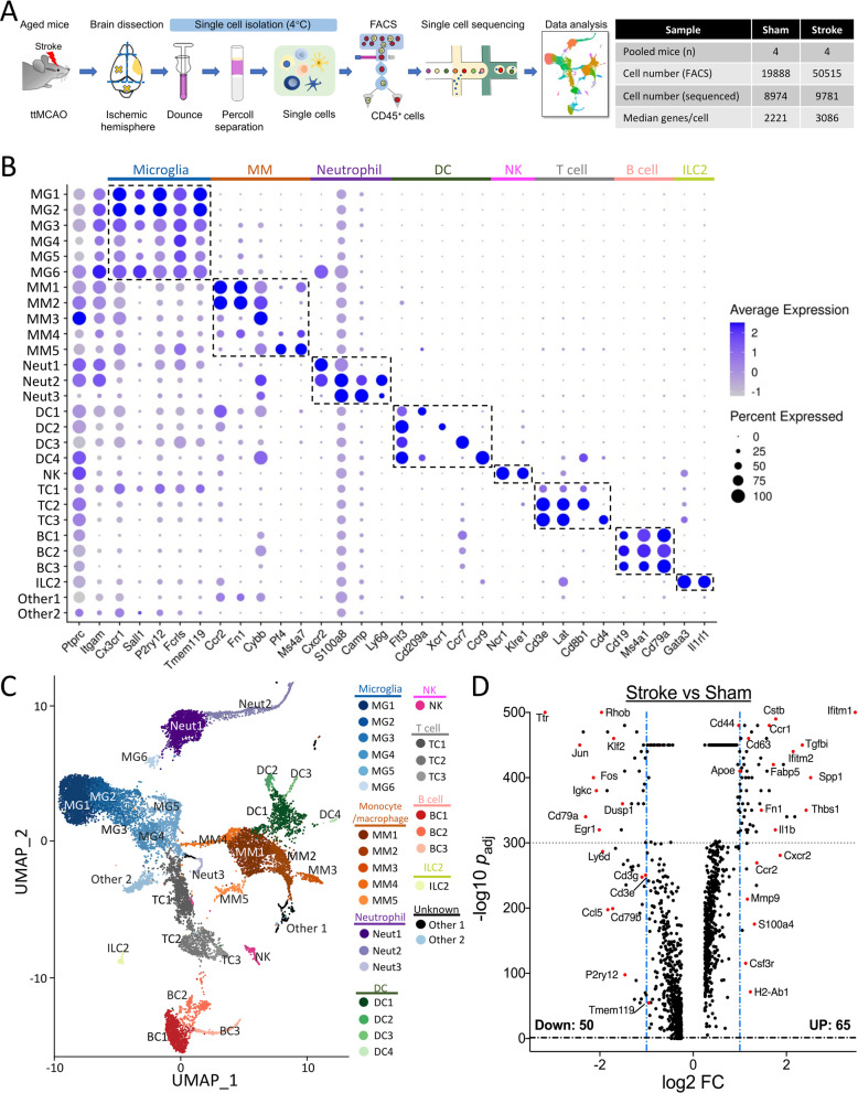
Fig. 2.
scRNA-seq analysis. A Workflow and data table of our scRNA-seq analysis. Aged mice were subjected to 6-h ttMCAO or sham. Three days later, both (sham) or ipsilesional (stroke) hemispheres were collected for scRNA-seq analysis. B Dot plot showing the scaled expression of selected signature genes for each cluster. Dot size depicts the percentage of cells within the cluster expressing each gene, and color intensity indicates the average expression level. C UMAP plot of aggregated data from both groups. D Volcano plot of differentially expressed genes (DEGs) between stroke and sham (Additional file 1: Table S4)

