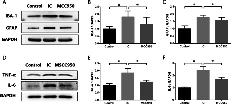
Fig. 2.
NLRP3 inflammasome contributes to activation of glial cells and process of neuroinflammation in SDH of IC rats. A–C Western blot analysis showing that expression levels of IBA-1 (microglia marker) and GFAP (astrocyte marker) were significantly increased in SDH of IC rats compared with normal rats, and MCC950 treatment significantly decreased expression levels of IBA-1 and GFAP in SDH of IC rats. D–F Western blot analysis showing that expression levels of proinflammatory cytokine TNF-α and IL-6 were significantly increased in SDH of IC rats compared with normal rats, and MCC950 treatment significantly decreased expression levels of TNF-α and IL-6 in SDH of IC rats. n = 8 per group. *P < 0.05

