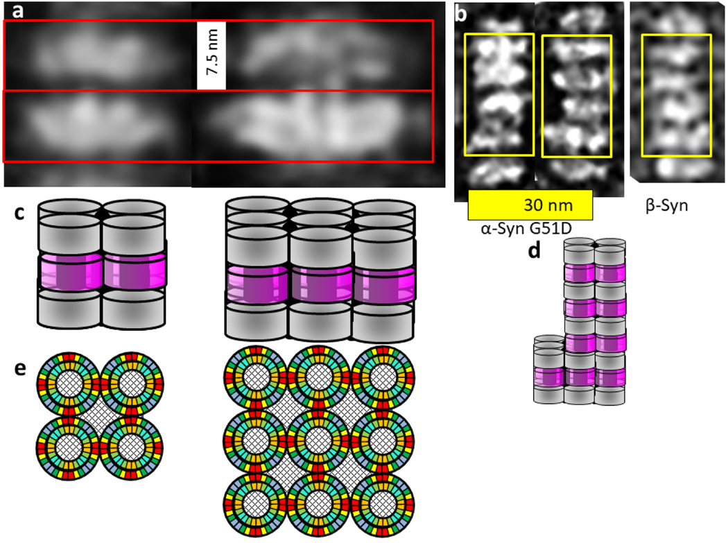
Figure 14.
Images and models of α-Syn and β-Syn stacked lipoprotein discs. (a) Two sizes of α-Syn stacked pairs obtained by image averaging. (b) Individual images of relatively long stacks for both α- and β-Syn. (Images modified from original EM images of Eichmann et al. [19]). (c) Side views of schematics of our models. The darkly shaded regions in the middle of and between the cylinders represent lipid-filled regions. The gray cylinders represent concentric β-barrels formed by the Nt and NAC domains from two discs; the magenta cylinders represent 32-stranded β-barrels formed by interlocking Ct domains from adjacent stacks. (d) A schematic model for the central stack illustrating variations of the numbers of octamers in adjacent discs of the stack above. (e) Wedge cross-sections representations of the Nt-NAC barrel regions of the octamers within a disc. The checkered regions inside and between the octamers represent lipid alkyl chains.

