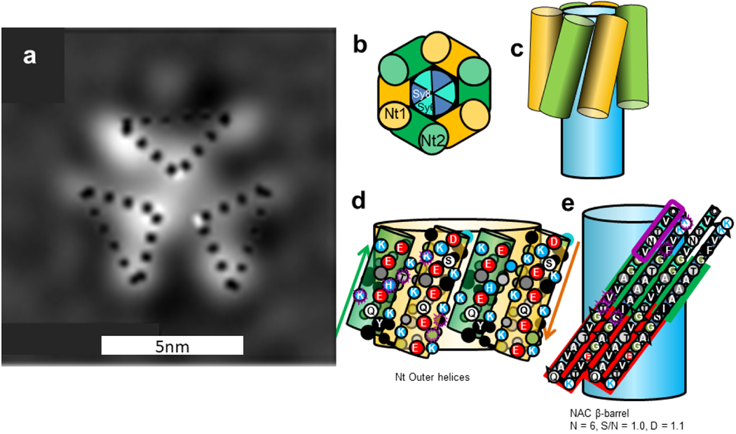
Figure 18.
(a) EM image of a predominately α-helical trimer (Copied from Wang et al. [41]) and (b-e) a Type 1 α-β barrel trimer model. This model resembles the tetramer of Fig. 5 except that there is one less subunit and all subunits are oriented in the same direction. (b) wedge representation, (c) side view schematic of helices, (d) side view of two subunits of the outer Nt helices, (e) flattened representation of two subunits of a core 6-stranded antiparallel β-barrel formed by three NAC β-hairpins. Residues in d and e that differ between α-Syn and β-Syn are enclosed in purple. The Sy7 has a red background.

