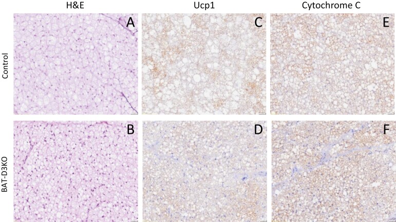
Figure 3.
Interscapular brown adipose tissue (iBAT) histology in adult BAT-D3KO mice. A and B, Representative microphotographs (×20 magnification) of sections stained with hematoxylin and eosin from control and BAT-D3KO mice as indicated; C and D, immunohistochemical image of BAT sections after staining with UCP1 antibody or with cytochrome-C antibody; E and F, images representative of 4 or 5 independent samples.

