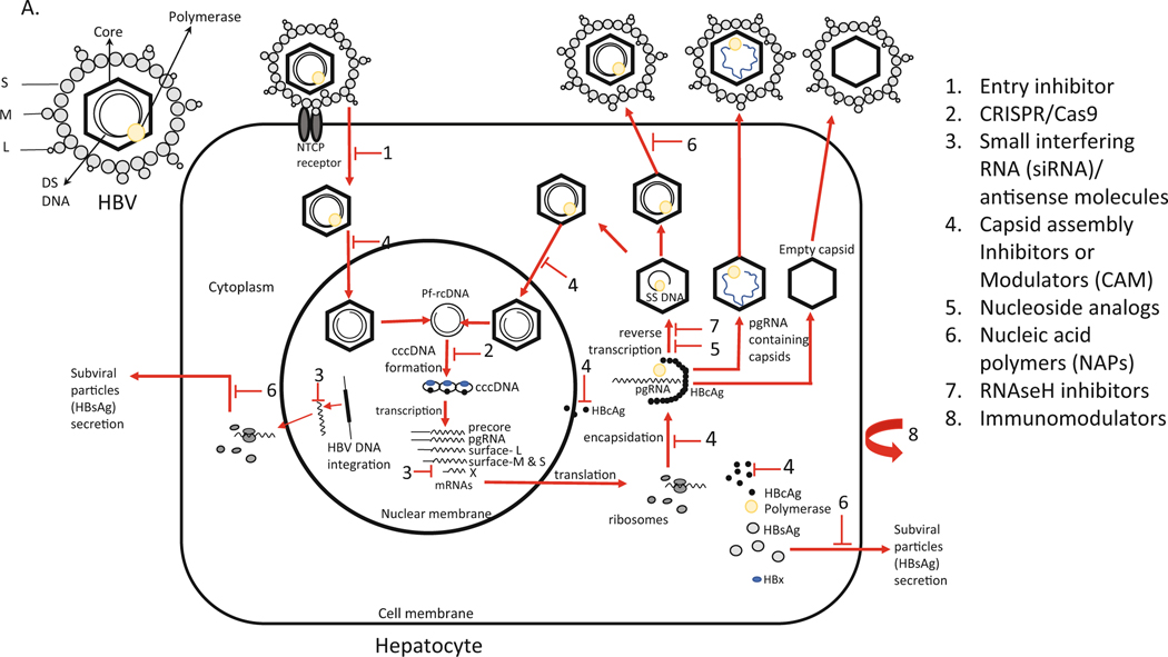
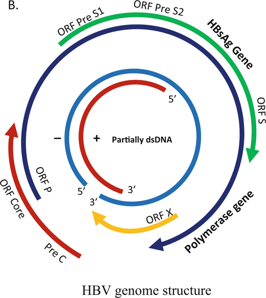
Fig. 5.1.
(a, b) HBV replication mechanism, genome structure, and schematic representation of inhibition sites. (a) The HBV has an envelope composed with three forms (large, middle, and small) of surface proteins that encloses the capsid with the double-stranded DNA genome. (b) Replication starts with HBV binding to the hepatocyte at the NTCP receptor. After entry, the viral particles are uncoated, and the nucleocapsid particle goes to the cellular nucleus. HBV protein free rcDNA (Pf-rcDNA) is converted to an episomal cccDNA, which is the transcription template for all four viral RNAs. The pgRNA is encapsidated together with viral polymerase and subsequently reverse-transcribed into viral minus strand DNA, followed by degradation of the RNA by RNAseH. Then, the plus-stranded DNA is synthesized to form the partially double-stranded relaxed circular DNA. Mature nucleocapsid can either be recycled back to the nucleus to maintain the pool of cccDNA or packed with envelope proteins and exported as infectious virions to infect other cells. pgRNA containing nucleocapsid and empty nucleocapsids are also packed with envelope proteins and released. S small, M medium, L large, DS double stranded, NTCP sodium taurocholate cotransporting polypeptide, CRISPR clustered regularly interspaced short palindrome repeats; CAS9 CRISPR associated 9


