Fig. 4.
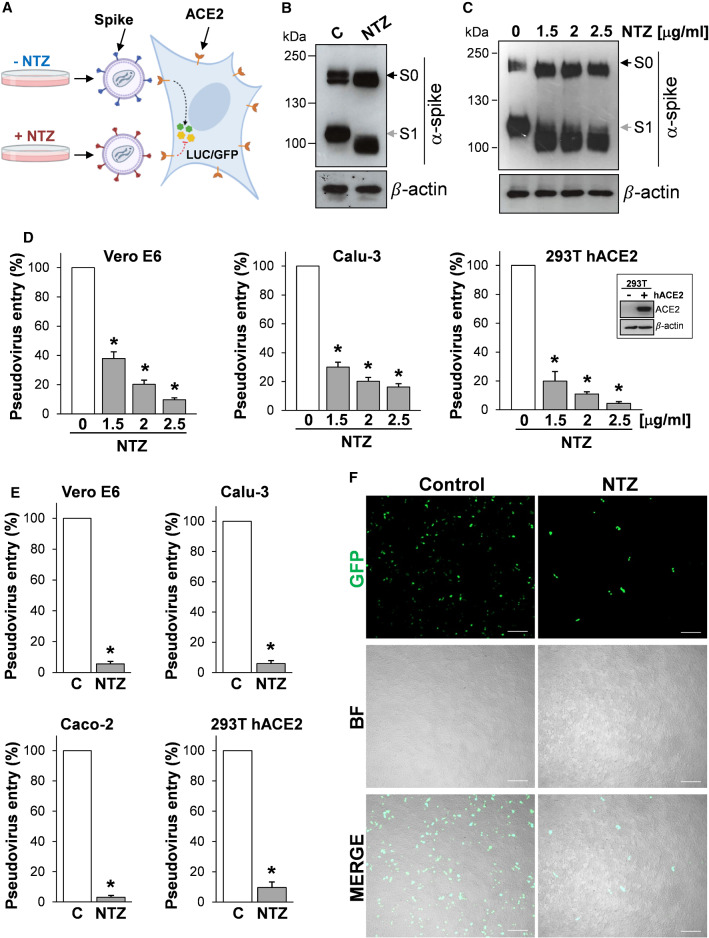
Nitazoxanide treatment inhibits SARS-CoV-2 S pseudovirus infectivity. A Diagram of SARS-CoV-2 pseudovirus luciferase (LUC) and green fluorescent protein (GFP) assay. SARS-CoV-2 spike-pseudotyped lentiviral (LV) or retroviral (MLV) particles carrying a reporter gene are produced in the presence of NTZ or vehicle (C) and used to infect different types of human cells, which will express LUC or GFP upon infection. B, C HEK293T cells transfected with plasmids for production of SARS-CoV-2 spike-pseudotyped-LUC LV particles (B) or SARS-CoV-2 spike-pseudotyped-LUC MLV particles (C) (see ‘Materials and methods’), were treated with 2.5 µg/ml (B) or different concentrations (C) of NTZ. At 48 h post-transfection, pseudovirus-containing supernatants were collected for infection of different types of cells (see panels D, E), whereas SARS-CoV-2 S expression was detected in WCE of HEK293T pseudovirus-producing cells by IB using anti-spike antibodies. D, E Vero E6, Calu-3, Caco-2 and hACE2-expressing HEK293T (293T hACE2) cells were infected with SARS-CoV-2 spike-pseudotyped MLV (D) or LV (E) particles obtained as described in B, C, and pseudovirus entry was analyzed by measuring LUC activities 72 h post-infection. hACE2 protein levels in HEK293T WT (−) and hACE2 (+) cells are shown in (D). Data are expressed as percent of untreated control. Error bars indicate means ± SD. *P < 0.05; ANOVA (D), Student’s t test (E). F SARS-CoV-2 spike-pseudotyped GFP lentiviral particles produced as described in (B) were used to infect 293T hACE2 cells. Pseudovirus-infected cells were visualized by fluorescence microscopy. Bright field (BF) and merge images are shown. Scale bar, 200 μm

