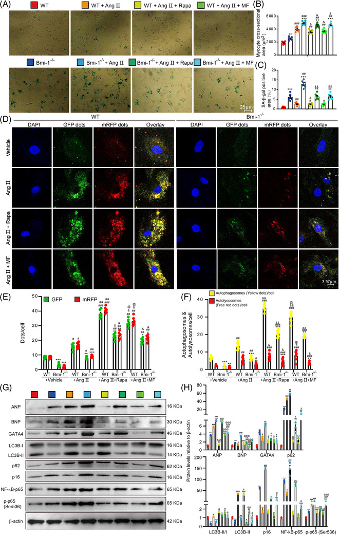
FIGURE 4.

Autophagy agonist supplementation improves GATA4‐dependent SA‐PCH of embryonic cardiomyocytes in WT mice more than Bmi‐1 –/– mice. Mouse embryonic cardiomyocytes (MECs) were isolated and cultured from 13.5‐day foetal hearts, and were treated with Ang II (5 × 10⁻⁶ mol/L for 70 h) to induce hypertrophy and were rescued with metformin (MF) (2.5 mM for 70 h) or rapamycin (Rapa) (100 nM for 70 h). (A) Representative micrographs of MECs stained for senescence‐associated β‐galactosidase (SA‐β‐gal). (B) Myocyte cross‐sectional area according to graph A. (C) The percentage of SA‐β‐gal‐positive area of myocytes. (D) Representative micrographs of WT and Bmi‐1–/– MECs, which transfected with autophagy lentivirus for 24 h under the treatment with Ang II together with metformin or rapamycin. (E) Percentage of positive GFP dots or mRFP dots relative to total cells in different treatment groups. (F) The number of autophagosomes and autolysosomes per cell. (G) Western blots of extracts from MECs showing ANP, BNP, GATA4, LC3B, p62, p16, NF‐κB‐p65 and p‐p65 (Ser536); β‐actin was the loading control. (H) Protein levels relative to β‐actin were assessed by densitometric analysis. The experiments were performed with three biological repetitions per group. Statistical analysis was performed with one‐way ANOVA test; values are mean ± SEM from six determinations per group, *p < .05, **p < .01, ***p < .001 compared to WT MECs with the same treatment; # p < .05, ## p < .01, ### p < .001 compared to control group with the same genotype; & p < .05, && p < .01, &&& p < .001 compared to Ang II‐treated group with the same genotype; @ p < .05, @@ p < .01, @@@ p < .001 compared to Ang II & Rapa‐treated group with the same genotype
