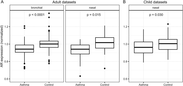
Figure 1.
Androgen receptor (AR) expression is lower in airway epithelial cells of asthma patients than control individuals. Box-and-whisker plots of normalized AR expression from bronchial (left) and nasal (right) epithelia in adults (A) and nasal epithelia in children (B). Center lines indicate median values, boxes indicate first quartile to third quartile range, whiskers indicate values up to 1.5 times interquartile range, and points indicate outlying values. For visual clarity, outliers below 0.6 normalized expression were excluded from the plot: one each from adult bronchial asthma, adult nasal asthma, and adult nasal control. Sample sizes for each group: A, bronchial asthma 264, control 146; nasal asthma 52, control 29; B, nasal asthma 49, control 58 (child data sets contained no bronchial samples). Adult data sets contained participant ages ranging from 18 to 74 years (mean, 35.7 years). Child data sets contained participant ages ranging from 6 to 16 years (mean, 11.0 years). P values from 2-tailed t tests of asthma vs control within each sample group are shown.

