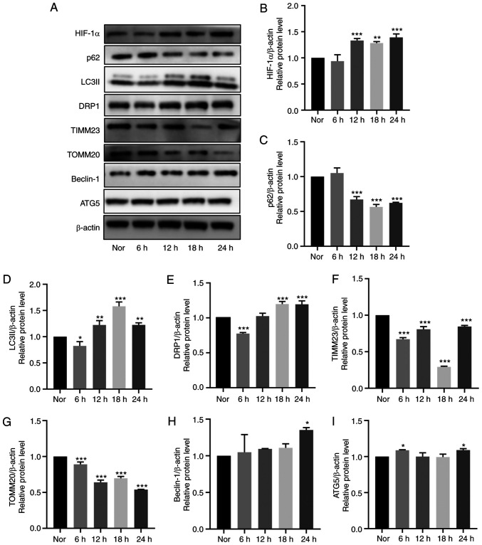
Figure 5.
Effects of hypoxia on mitophagy in cultured HDPCs. (A) Representative western blotting images. Protein expression of (B) HIF-1α, (C) p62, (D) LC3II, (E) DRP1, (F) TIMM23, (G) TOMM20, (H) Beclin-1 and (I) ATG5 in HDPCs cultured in hypoxia were quantified. The expression of the target proteins was measured by quantifying the intensity of the bands and normalized to that of β-actin. The results are presented as the means ± SD from ≥ three independent experiments. *P<0.05, **P<0.01 and ***P<0.001 vs. normoxia. Nor, normoxia; HIF-1α, hypoxia-inducible factor-1α; DRP1, dynamin-related protein 1; TIMM23, translocase of inner mitochondrial membrane 23; TOMM20, translocase of outer mitochondrial membrane 20; ATG5, autophagy related 5.

