
Figure 3.
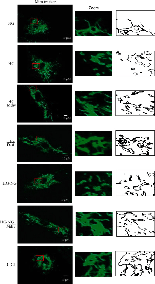
Direct inhibition of Drp1 during reversal phase prevents mitochondrial fragmentation. Mitochondrial morphology was evaluated by live cell microscopy (63x) using MitoTracker Green dye. The middle panel displays the area inside the box, and the right panel shows the analysis of the area inside the box by ImageJ for particles (size = 0‐infinity; circularity = 0 to 1) after adjusting for color threshold. Bare outlines were drawn to illustrate the morphology. Each image is representative of 5-7 cells/group, repeated in 2-3 different cell preparations.