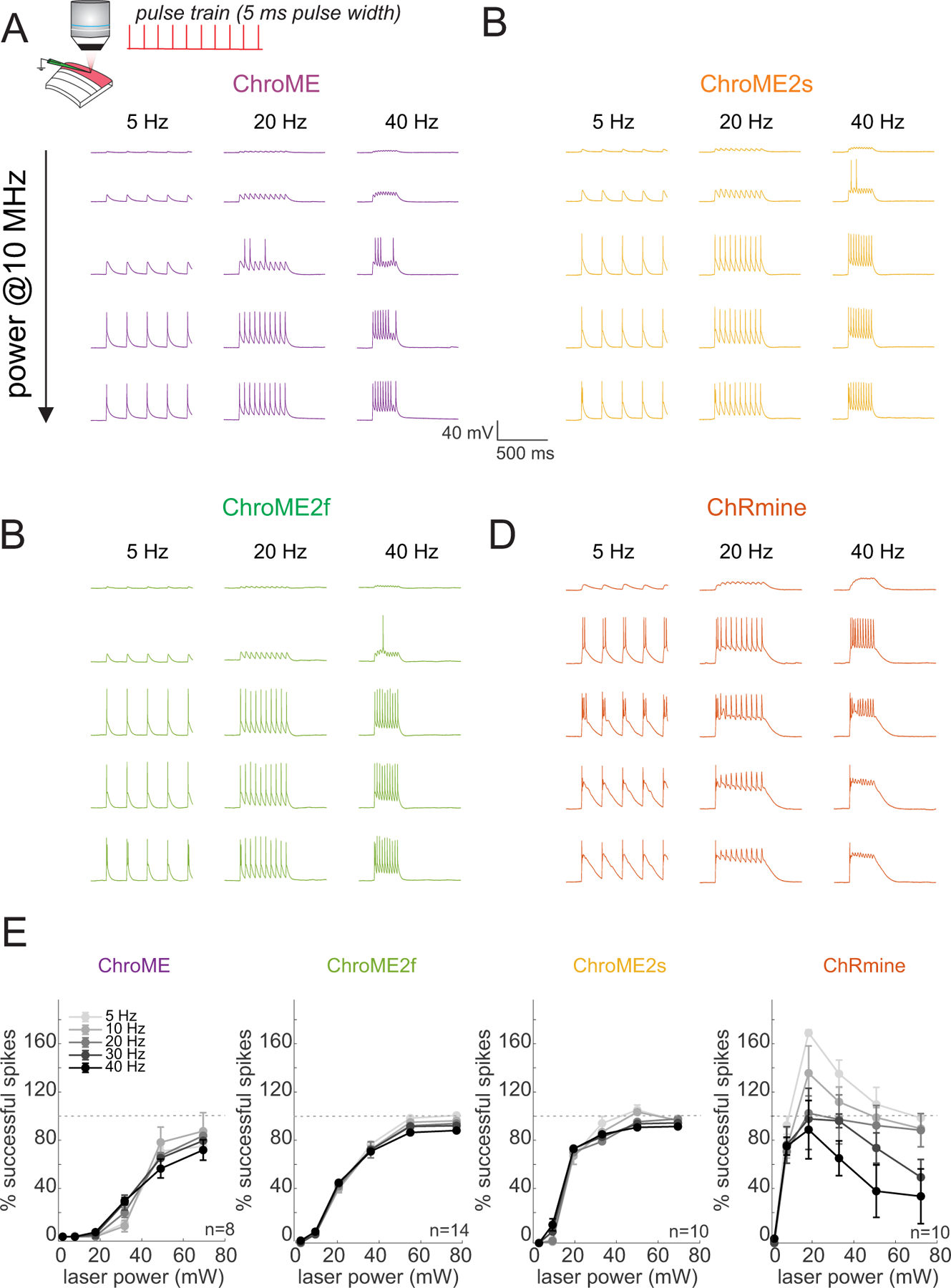
Figure 3. Comparison of two-photon light-evoked spiking of L2/3 pyramidal neurons expressing ChroME2.0 variants to ChroME and ChRmine.

A) Top: Schematic the experiment. A single L2/3 neuron is patched in current clamp mode and illuminated with fixed frequency trains of 5 ms pulses at 1040 nm, 10 MHz repetition rate. Bottom: example traces from a ChroME-expressing neuron,
B-D) Example traces from representative ChroME2f, ChroME2s, and ChRmine-expressing cells.
E) Plots of the fraction of 5 ms light pulses that drove spikes across laser powers and stimulation frequencies for the four opsins. Sample size (cells) is indicated in the panels. The two-photon excitation spot size was 12.5 um in diameter. Data represent the mean ± s.e.m. ChroME: n=8 cells, 1 mouse; ChroME2f: n =14 cells, 2 mice; ChroME2s: n = 10 cells, n = 1 mouse; ChRmine: n = 10 cells, 2 mice. >100% successful spikes indicates more than one spike generated per light pulse (‘extra spikes’).
