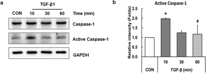
Fig. 3.
Treatment of TGF-β activates caspase-1 in LX-2 cells. LX-2 cells were treated with 10 ng/ml TGF-β for 10, 30, or 60 min. a Cell lysates were prepared, and protein expression was analyzed by western blotting. GAPDH was used as an internal control. b Relative levels of active caspase-1 were normalized to GAPDH and quantified using ImageJ software. Data are means ± SEM. *p < 0.05 vs. CON; #p < 0.05 vs. TGF-β (10 min) (n = 3/group)

