. 2022 Mar 24;34(2):110–117. doi: 10.5021/ad.2022.34.2.110

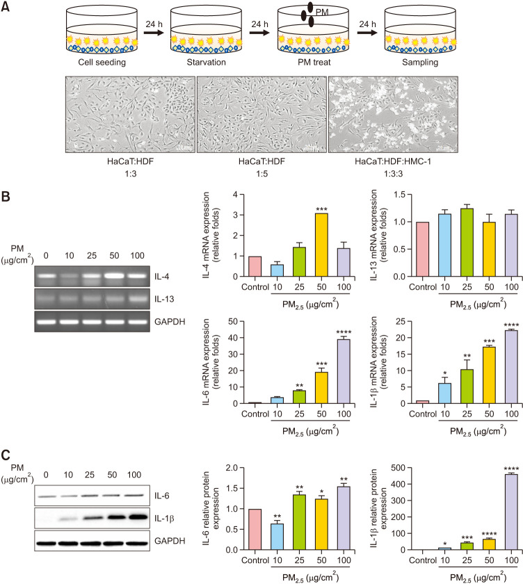
Fig. 1. Particulate matter (PM)-induced proinflammatory responses in the triple-cell culture model. (A) Schematic representation of the strategy of PM exposure in the triple-cell culture model. Morphology of co-cultured keratinocytes, fibroblasts, and mast cells examined using phase-contrast microscopy. The cells were serum-starved overnight and treated with PM (0, 10, 25, 50, and 100 µg/cm2) for 24 hours. (B) The mRNA expression levels of interleukin (IL)-4, IL-13, IL-6, and IL-1β are presented in a graphical form (the relative mRNA levels are presented as fold change compared with untreated cells). (C) The protein expression levels of IL-6 and IL-1β were determined using western blotting. Quantification of protein levels. Values are presented as mean±standard deviation (n=3). *p<0.05, **p<0.01, ***p<0.001, ****p<0.0001 compared with basal levels.

Fig. 1