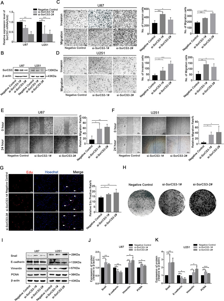
Fig. 3. Knockdown of SorCS3 promotes the proliferation, migration and invasion of glioma cells in vitro.
A, B After siRNA-SorCS3-1# and 2# transfection, the expression levels of SorCS3 were examined through real-time PCR and western blotting, using β-actin as an endogenous control. C, D Transwell assays used to determine the influence of transfection with si-SorCS3-1# and 2# on the migratory and invasive abilities of U87 and U251 cells. E, F Effect of SorCS3 knockdown on wound healing of U87 and U251 cells. G Cell proliferation was determined by EdU staining in SorCS3 knockdown condition and EdU incorporation was calculated as EdU+ cells/total cells, quantified by ImageJ. Red was stained for proliferation (EdU + ), blue was stained for nucleus. H Colony formation assay for assessing the cell proliferation of knockdown SorCS3. I–K Western blot analysis of proliferation- and EMT-associated marker after transfection with SorCS3 siRNA. Data are shown as mean ± S.D. including three independent experiments. *p < 0.05; **p < 0.01; ***p < 0.001.

