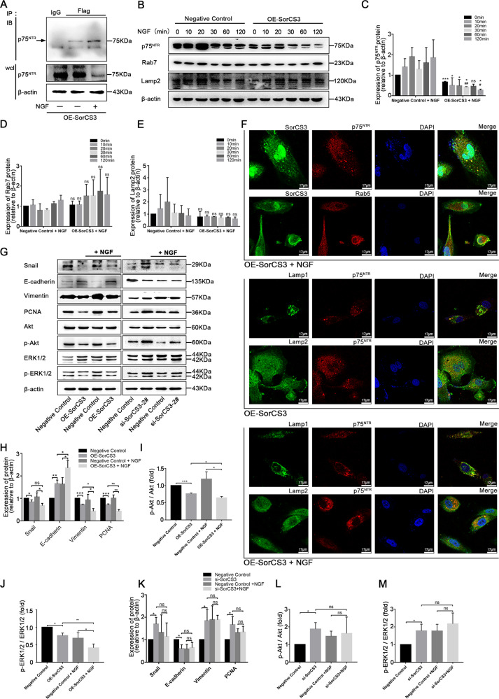
Fig. 5. NGF promotes the SorCS3 and p75NTR interaction.
A U87 cells were transfected SorCS3-Flag plasmid, and then stimulated or not with NGF (100 ng/mL) for 60 min. IP were performed using anti-Flag antibody, and the immunocomplexes were immunoblotted (IB) using anti-p75NTR antibody. In parallel, immunoblots for p75NTR were performed on whole-cell lysates (wcl); the isotypic lane Immunoglobulin G (IgG) represents the IP control. B–E U87 overexpression of SorCS3 and control were stimulated with NGF (100 ng/mL) over 0–120 min time course. Cell lysates were analyzed by western blotting for components of the canonical internalization signaling pathway using the indicated antibodies. *p vs. the control group, #p vs. the OE-SorCS3 (NGF, 0 min) group. ns: not statistically significant. F Process of internalization was visualized by immunofluorescence staining of the endosome and the lysosomal marker. U87 cells were stimulated with NGF (100 ng/mL) for 60 min, and then immunofluorescence for SorCS3-Flag, p75NTR, early endosome markers (Rab5) and lysosome markers (Lamp1 and Lamp2). Scale bar: 17 µm. G–M U87 were transfected with SorCS3-Flag plasmid or siRNA (si-SorCS3-2#) and the corresponding control, and then stimulated with NGF (100 ng/mL) for 60 min. Cell lysates were analyzed by western blotting with the indicated antibodies, using β-actin as an endogenous control. Data are shown as mean ± S.D. including three independent experiments. *p < 0.05; **p < 0.01; ***p < 0.001.

