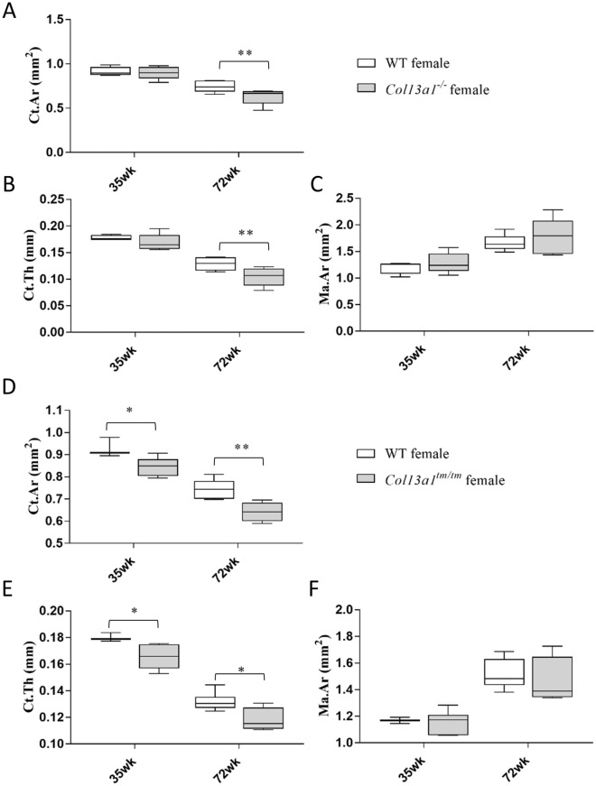
Figure 6.
Morphometric parameters for HRCT analysis of metaphyseal cortical bone. The Col13a1−/− female mice and respective WT controls (A–C). There were no statistically significant differences in the cortical area (Ct.Ar, A) or thickness (Ct.Th, B) or the medullary area (Ma.Ar, C) at the 35-week time point. At the 72-week time point, Ct.Ar and Ct.Th were significantly reduced in the Col13a1−/− samples compared to the WT mice. The Col13a1tm/tm female mice and respective WT controls (D–F). The cortical area (Ct.Ar, D) and thickness (Ct.Th, E) were significantly reduced in the Col13a1tm/tm mice at the 35- and 72-week time points compared to the respective controls. The medullary area (Ma.Ar, F) was unaltered. n(WT female): 4–5; n(Col13a1−/− female): 5; n(WT female): 3–6; n(Col13a1tm/tm female): 4–6; the whiskers represent min to max; **q < 0.01 determined by two-way ANOVA and followed by the false discovery rate.

