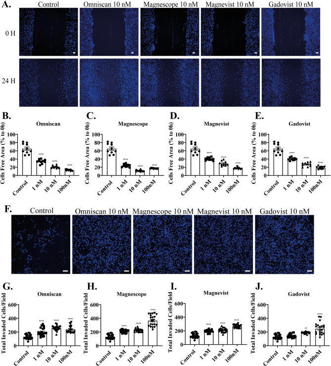
Figure 2.
GBCAs accelerated astrocyte migration. (A) Representative photomicrographs showing the effects of GBCAs on 2D wound healing assays using C6 cells. Live-cell staining was performed using Cellstain-Hoechst 33,258 (Dojindo Molecular Technologies, Inc., Japan). Quantitative analysis of the effect of Omniscan (B), Magnescope (C), Magnevist (D), and Gadovist (E) (1–100 nM) on cell migration measured by wound healing assay. (F) Representative photomicrographs showing the effects of GBCAs on 3D matrigel invasion assays using astrocytes. Cell nuclei were stained with DAPI. Quantitative analysis of the effect of Omniscan (G), Magnescope (H), Magnevist (I), and Gadovist (J) (1–100 nM) on cell invasion was performed by matrigel invasion assay. The total number of cells was quantified using ImageJ software (NIH). Bars represent 50 μm. Data are expressed as the mean ± SEM of at least three independent experiments. ****p < 0.0001, **p < 0.01, indicates statistical significance were analysed by ANOVA continued with post hoc Bonferroni’s or Turkey test compared with the control.

