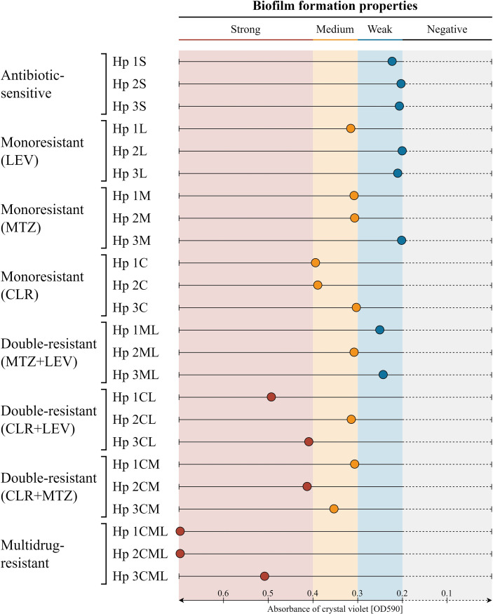
Figure 1.
Graphical representation of the biofilm formation of 24 clinical H. pylori strains in static conditions and a 3-day incubation, determined using a crystal violet staining method. The H. pylori strains were classified as strong, medium and weak biofilm producers when the OD590 of the crystal violet solution was equal to ≥ 0.4, 0.4 - 0.3, 0.3 - 0.2, respectively. Values obtained for the pure medium, being a negative control, were < 0.2 and indicated the lack of biofilm production by the tested strain. The tests were performed in three biological replications with six technical repetitions (n = 18/strain). CLR, clarithromycin; MTZ, metronidazole; LEV, levofloxacin.

