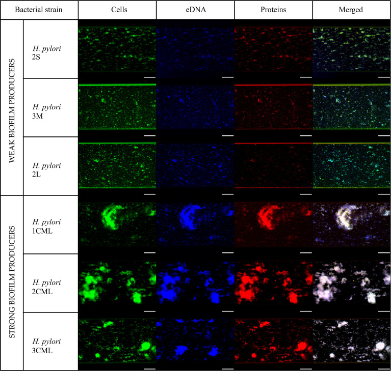
Figure 3.
Representative fluorescence microscopy photographs of biofilms produced by six selected H. pylori strains incubated for 24 h under microfluidic conditions. Biofilms were stained by SYTO9 (green), DAPI (blue) and SYPRO RUBY (red) to enable determination of the bacterial biomass, extracellular DNA and extracellular proteins of the biofilm matrix, respectively. Scale bars show 20 µm.

