
FIGURE 1.
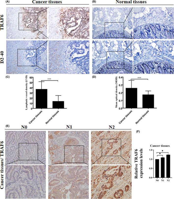
Expression of TRAF6 and lymphatic vessels in colorectal cancer clinical samples. (A, B) Representative immunohistochemical staining images for TRAF6 protein and D2‐40 in colorectal cancer (CRC) and adjacent normal tissues from a tissue array are shown. Magnification, ×40 and ×100. (C) Mean optical density (MOD) showed that TRAF6 expression in colorectal cancer tissues was elevated compared with normal colorectal tissues. (D) The result of LVD showed that the expression of lymphatic vessels in colorectal cancer tissues was elevated compared with normal colorectal tissues. (E) Representative immunohistochemical staining images for TRAF6 protein in CRC tissues from a tissue array were shown. Magnification, ×100 and ×200. (F) The result of immunohistochemical staining showed that the expression of relative TRAF6 expression levels in N1 and N2 stage colorectal cancer tissues was elevated compared with N0. Data are shown as mean ±SD. ***P < 0.001