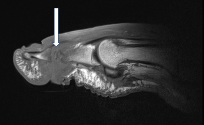
Figure 1.
T1-weighted sagittal images of a right first interphalangeal joint demonstrating osteomyelitis of the head of the proximal phalanx and the basis of the distal phalanx. Infectious osteomyelitis is indicated by fat mark suppression in T1 (arrow). Magnetic resonance images with consent of the patient.

