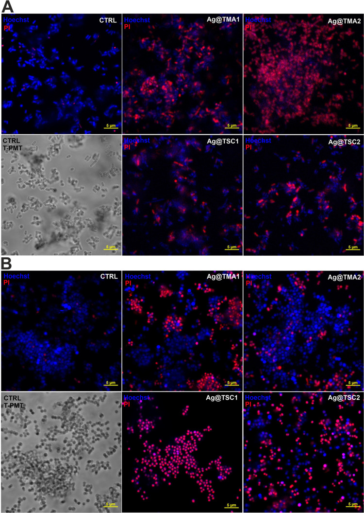
Figure 6.
Laser scanning confocal microscopy imaging of live/dead staining of bacteria after 2 h of incubation with AgNPs: (A)E. coli and (B) S. aureus. The live cells were stained with Hoechst33342 (blue fluorescence), and dead cells were stained with propidium iodide (PI, red fluorescence). Scale bar—5 μm.

