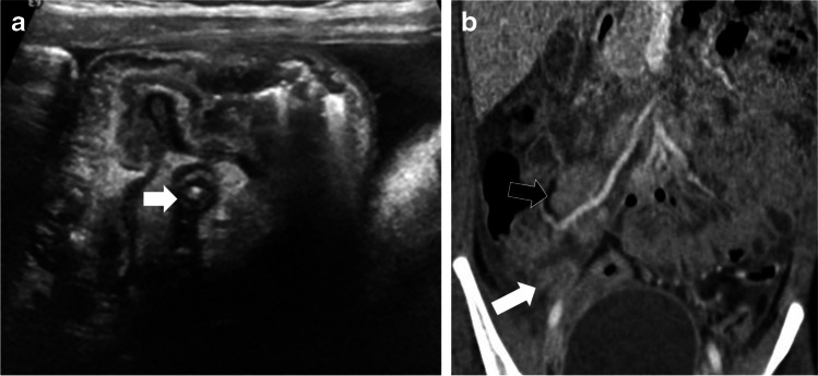
Fig. 2.
Case 4: An 8-year-old girl presented with abdominal pain, fever, and vomiting. a Initial transverse US section of right iliac fossa on presentation shows inflamed appendix (arrow) with inflammatory changes. Findings were suspicious for appendicitis. b CT scan (coronal post-contrast) shows multiple enlarged ileocolic lymph nodes (black arrow), largest 20 mm in short axis, free fluid in right iliac fossa, inflamed mesentery, ileal and cecal thickening and inflamed appendix (8 mm, white arrow)

