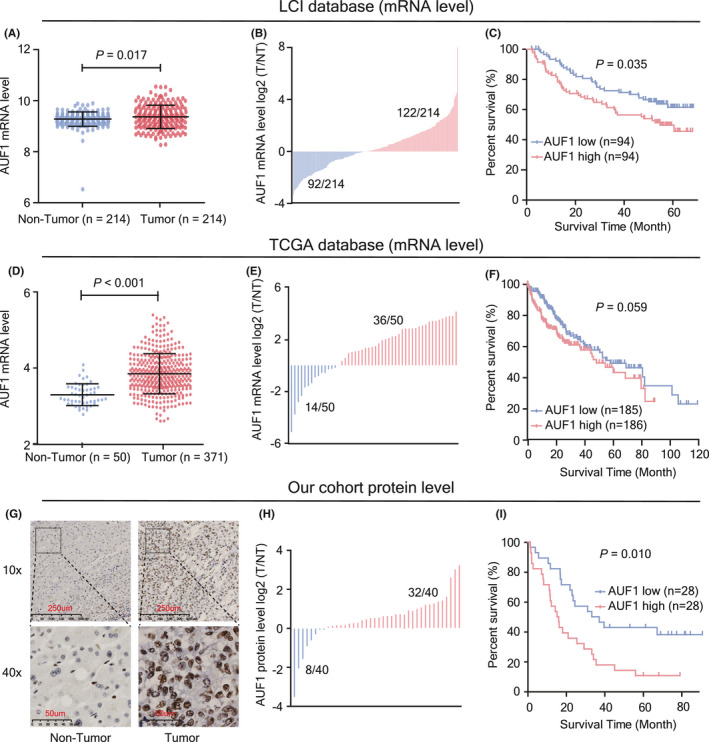
FIGURE 1.

Expression and prognostic value of AUF1 in patients with HCC. A, AUF1 mRNA levels in HCC tissues and matched non‐tumor tissues from the LCI database (n = 214). B, Abnormal expression of AUF1 mRNA in 214 HCC tissues from the LCI database. T/NT indicates the expression level ratio of tumor tissues and their corresponding non‐tumor tissues. C, Kaplan‐Meier analysis of AUF1 mRNA and overall survival from 188 HBV‐HCC tissues from the LCI database. D, AUF1 mRNA level in 371 HCC tissues and 50 non‐tumor tissues from TCGA database. E, Abnormal expression of AUF1 mRNA in 50 HCC tissues from TCGA database. F, Kaplan‐Meier analysis of AUF1 mRNA and overall survival from 371 HCC tissues from TCGA database. G, H, AUF1 protein levels in paired HBV‐HCC tissues detected using immunohistochemistry (IHC) staining (n = 66) and western blot assay (n = 40). I, Kaplan‐Meier analysis of AUF1 IHC score and overall survival from 56 HBV‐HCC tissues
