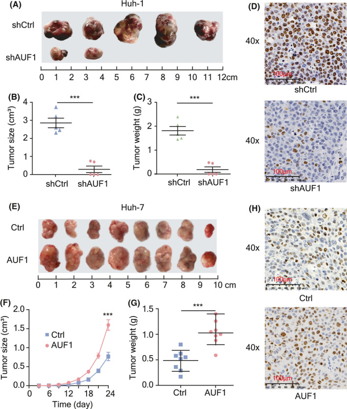
FIGURE 3.

AUF1 promotes HCC tumorigenicity in vivo. A‐C, Subcutaneous injection of Huh‐1 cells with stable knockdown of AUF1 or control cells into NPG/Vst mice (n = 5). Images of neoplasms from each group of NPG/Vst mice (A), measurement of tumor volumes (B), and tumor weight (C). D, Immunohistochemistry staining of proliferation marker Ki‐67 in tumor tissues. Scale bars, 100 μm. E‐G, Subcutaneous tumor model of Huh‐7 cells with AUF1 overexpression or control cells injected into NPG/Vst mice (n = 8). Images of neoplasms from each group of NPG/Vst mice (E), measurement of tumor volumes (F), and tumor weight (G). H, Immunohistochemistry staining images of Ki67in tumor tissues. Scale bars, 100 μm. ***P < .001
