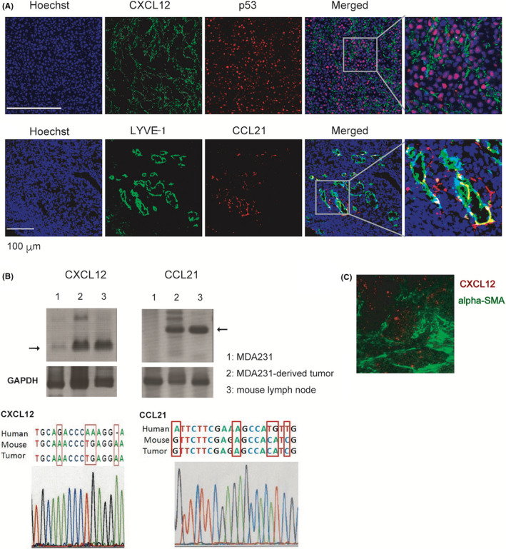
FIGURE 3.

Expression of CXCL12 and CCL21 in MDA231‐derived tumor. A, Parental MDA231 cells (1 × 10⁶) were injected into the thoracic mammary fat pad of female SCID mice at 9 weeks of age (n = 3). A sham operation was performed on the contralateral side. Twelve weeks after injection, primary tumors were dissected. Serial sections of fresh‐frozen tumor specimens from tumor xenograft were subjected to immunohistochemical staining with anti‐CXCL12 (green, upper panel) and anti‐human p53 mAb (red, upper panel) or anti‐LYVE‐1 (green, lower panel) and anti‐CCL21 (red, lower panel) antibodies. Scale bar, 100 µm. B, Upper row: RT‐PCR analysis of CXCL12 and CCL21 in the parental MDA231 cell line, MDA231‐derived tumor tissue, and C57BL/6 mouse lymph node using oligonucleotide primer pairs common to mouse and human sequences. GAPDH were used as an endogenous control. Bottom: the tumor‐derived nucleotide sequence of the RT‐PCR products was determined and compared with the species‐specific nucleotide positions of the CXCL12 and CCL21 genes. C, Localization of CXCL12 expression (red) and alpha‐smooth muscle actin (α‐SMA)‐positive cancer‐associated fibroblast (green) in MDA231‐derived tumor tissue
