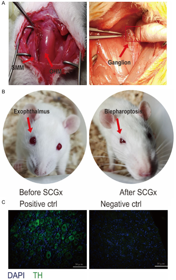
Figure 1.

Surgical removal of the SCG in rats and verification by performance. A. The anatomy of the SCG and the neighboring tissues. The glandular tissue and sternomastoid muscle (SMM) were dissected bluntly to make clear the CCA. The SCG was situated behind the carotid bifurcation, and the cell body of the ganglion was gently pulled until their full avulsion from the sympathetic chain. B. The blephar before and after bilateral superior cervical ganglionectomy. C. Immunofluorescence staining of SCG. The same staining protocol was used as a negative control, except that the TH antibody was applied. Tyrosine hydroxylase (TH), scale bar: 50 μm.
