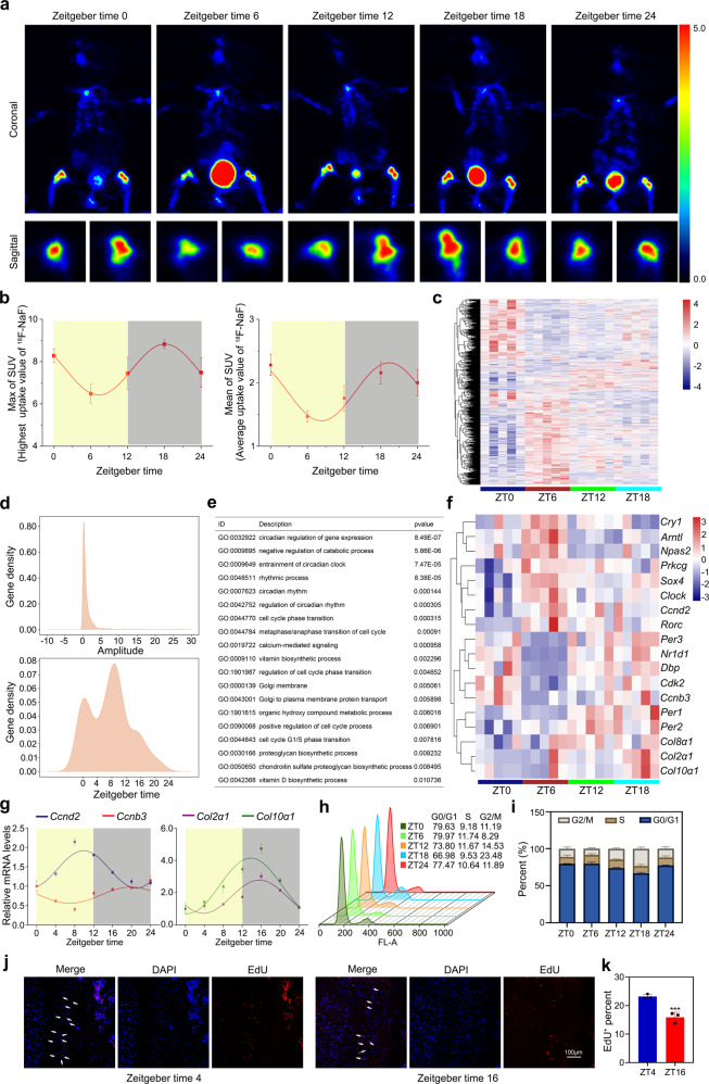
Fig. 1. Chondrification center possesses a circadian rhythm of cell proliferation and matrix synthesis.
a Positron emission tomography (PET) scans of C57BL/6J mice after 18F-labeled sodium fluoride (18F-NaF) tail vein injection at indicated times (n = 3 per group). b PET analysis of max and mean SUV. c Heatmaps representing expression levels of circadian genes in the indicated genotypes. d The gene density of amplitude (upper) or phase (under) distribution of all circadian oscillation genes in wild-type (WT) mice. e Associated Gene Ontology (GO) across all circadian genes in WT. f Heatmap showed some circadian genes that were related to circadian rhythm, cell cycle, and collagen synthesis. g qRT-PCR analysis of the diurnal rhythms of Ccnd2, Ccnb3, Col2α1, and Col10α1 in the growth plate cartilages of distal femora at indicated times (n = 3 for each bar). h, i Cell-cycle distribution of the growth plate cartilages tissue determined by the flow cytometry (n = 3 for each bar). j, k EdU staining of proliferating cells in proliferation zone (PZ) from the growth plate cartilages at ZT4 and ZT16. Arrowheads indicate EdU+ cells, ***P < 0.001 (n = 3 for each bar). Scale bar, 100 μm.

