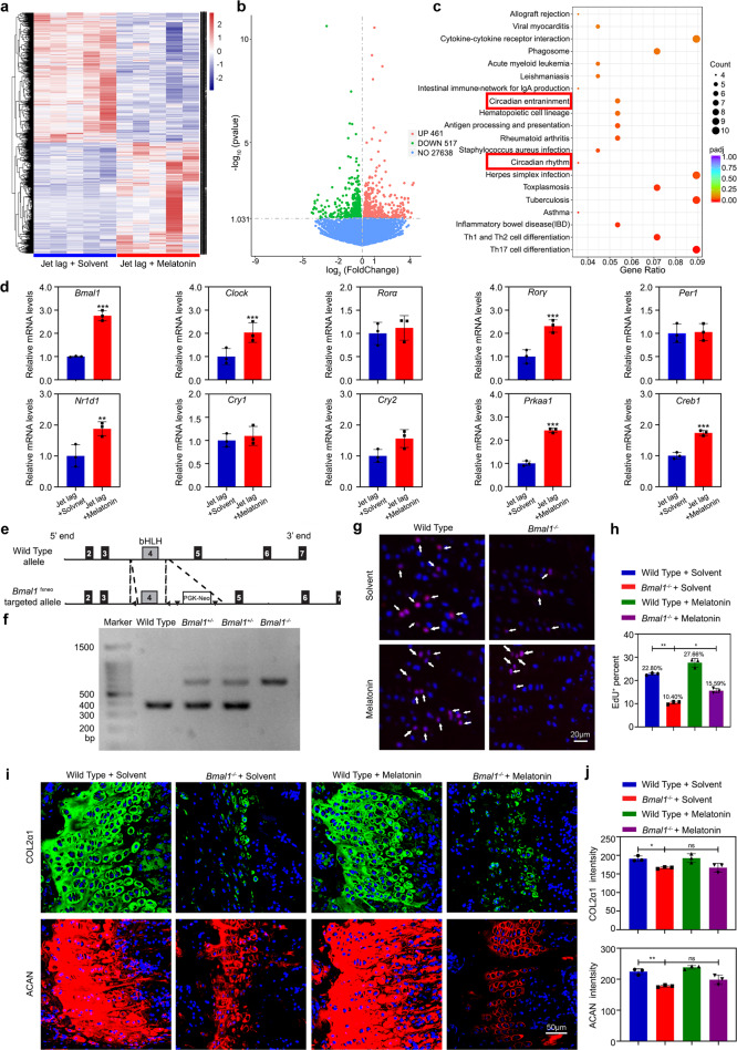
Fig. 6. BMAL1 acts as a principal molecule in circadian cell proliferation and matrix synthesis.
a Heatmap of differentially expressed genes (DEGs) in jet-lagged mice treated with equal solvent or melatonin (n = 5 pairs per group). b Volcano plot of jet-lagged mice treated with equal solvent or melatonin. c Associated Gene Ontology (GO) across jet-lagged mice treated with equal solvent and melatonin. d Confirmation of the DEGs in the growth plate tissue from jet-lagged mice treated with equal solvent or melatonin by qRT-PCR (n = 3 for each bar). Data represented as mean ± SD, **P < 0.01, ***P < 0.001. e, f The genotypes of WT, Bmal1+/-, and Bmal1-/- mice analysis by PCR. g, h EdU staining of proliferating cells in PZ from the growth plate cartilages in wild-type and Bmal1-/- mice with melatonin or equal solvent. Arrowheads indicate EdU+ cells (n = 3 for each bar). Scale bar, 20 μm. i, j COL2α1 and ACAN immunofluorescence images and quantitative analysis of labeling intensity in the growth plate cartilages from wild-type and Bmal1-/- mice with melatonin or equal solvent at ZT10 (n = 3 for each bar). *P < 0.05, **P < 0.01, ns, nonsignificant. Data were analyzed using one-way ANOVA with Tukey multiple comparisons test. Scale bar, 50 μm. (Sigma, M5250, 5 mg/kg/d, Bmal1-/- mice were injected from 3 weeks of age, injected before the dark onset for 28 days).

