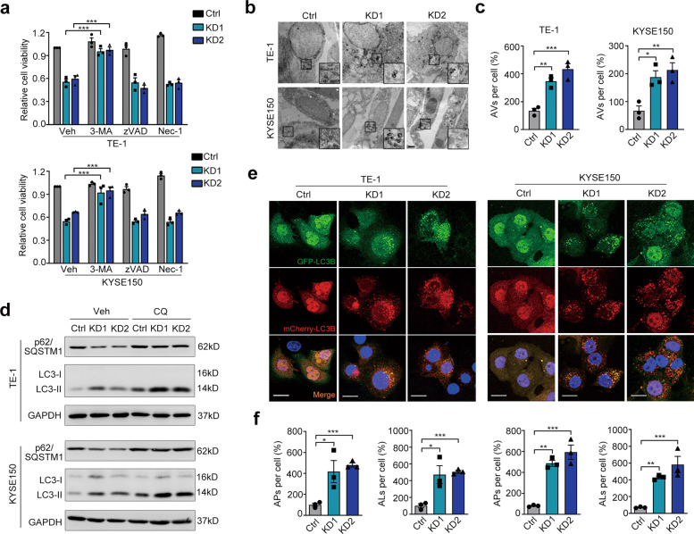
Fig. 2. Autophagic cell death is involved in LOX-1-knockdown mediated tumor inhibition.
a TE-1 and KYSE150 cells transfected with LOX-1 siRNA or control were treated with 3-methyladenine (3-MA, 200 µM) or Z-VAD-FMK (zVAD, 10 µM) or necrosis inhibitor Nec-1 (10 µM), and cell viability was detected by CCK-8 assay (Student’s t-test). b Ultrastructure of cells transfected with LOX-1 siRNA or control were analyzed by transmission electron microscopy (TEM) (Scale bar, 2 µm). c Statistic analysis of autophagic vacuoles (AVs) induced by LOX-1 knockdown (Student’s t-test). d Cells transfected with LOX-1 siRNA or control were treated with lysosome inhibitor chloroquine (CQ, 50 µM) or vehicle (Veh) for 4 h, and the changes of autophagy markers were analyzed by immunoblot. e Autophagic flux detection with the mCherry-GFP-LC3 reporter. Cells stably expressing mCherry-GFP-LC3 were transfected with LOX-1 siRNA or control, confocal microscopy images of representative cells were acquired (Scale bars, 20 µm). Blue represents DAPI; APs, mCherry+ autophagosomes; ALs, mCherry+ GFP− autolysosomes. f The percentages of vesicles per cell were calculated (Student’s t-test). *P < 0.05, **P < 0.01, ***P < 0.001.

