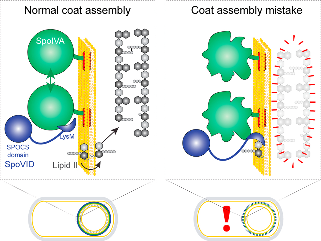
Figure 7. Model for the SpoVID-mediated orchestrated assembly of coat and cortex during sporulation.
Depicted are a wild-type sporulating bacterium (left) and a mutant bacterium that mis-assembles the spore coat (right). An expansion of the spore envelope (spore coat basement layer, outer forespore membrane, and cortex) is depicted above each cell. When SpoIVA (green) assembles properly in a wildtype cell, the LysM domain of SpoVID (blue) is occluded. Lipid II (gray) is produced in the mother cell and is permitted to flip to the intermembrane space to incorporate into the growing cortex. When SpoIVA mis-assembles (right), the LysM domain of SpoVID is liberated and sequesters lipid II, preventing it from being flipped into the intermembrane space and thereby disrupting cortex assembly.

