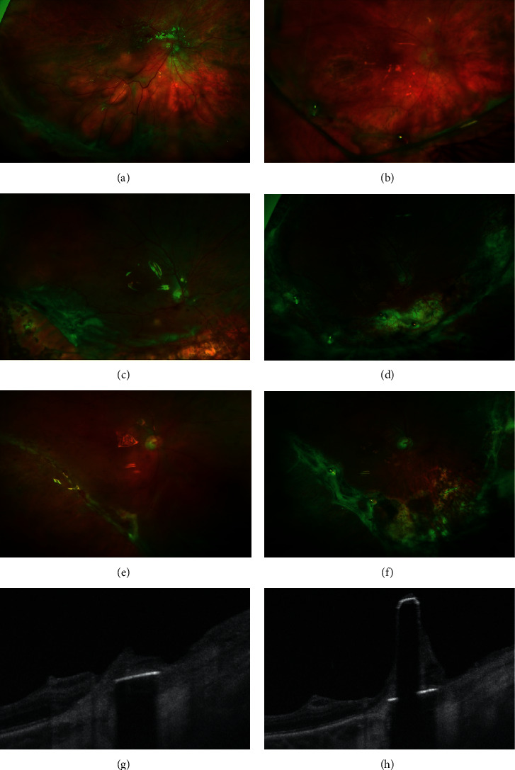
Figure 2.

Ultra-widefield scanning laser ophthalmoscopy. Fundus photography of patient 1 before surgery (a) and 5 weeks after surgery (b). 2 retinal tacks (RT) are placed at 6 o'clock and 7 : 30 positions. A fibrous reaction at the RT situs and along the retinectomy is seen. The retinectomy borders are not detached, and the fibrous strands are held by RTs. Fundus photography of patient 3 before surgery (c) and 6 months after surgery (d). 4 RTs are placed at 5 : 30, 6, 7, and 8 o'clock positions. A fibrous reaction involves RTs and retinectomy borders. Fundus photography of patient 8 before surgery (e) and 11 months after surgery (f). 2 retinal RTs were placed at 6 : 30 and 8 : 30 positions to reattach the stiff retina of the retinectomy border close to the macula. Retinectomy borders are not everted after surgery, despite fibrous reaction (b, d, f). Swept-source optical coherence tomography (SS-OCT) scans with segmentation of the RT in patient 11 (g, h). SS-OCT of the RT shows a fibrous reaction around the tack and on the tack's shaft.
