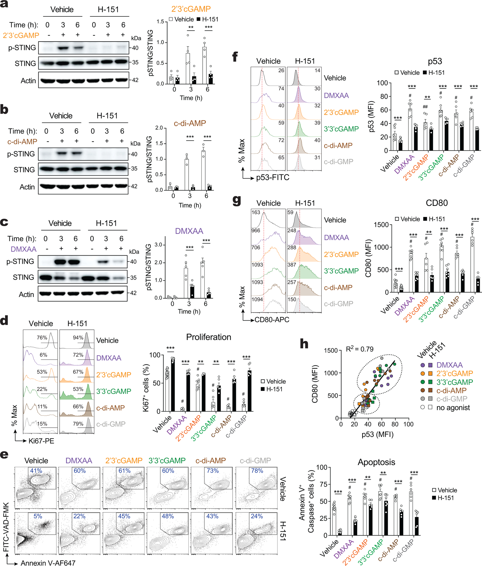
Extended Data Figure 8. Inhibition of STING increases proliferation and decreases apoptosis, p53 and CD80 expression in T cells.

(a-c) Immunoblots of total and phospho-STING (S366) protein expression in WT CD4+ T cells activated for 2 days with anti-CD3+28 and treated or not with the STING inhibitor H-151. Cell were stimulated with 10 μg/ml 2’3’cGAMP (a), 5 μg/ml c-di-AMP (b) or 3 μg/ml DMXAA (c) for 3–6 hours. Actin was used as loading control. Representative blots (left) and quantification (right) of at least 3 independent experiments and 4, 3, and 5 mice for 2’3’cGAMP, c-di-AMP, and DMXAA treatment, respectively. (d-g) Flow cytometry analysis of Ki67 expression (d), apoptosis measured by annexin V and active caspase (e), p53 (f), and CD80 expression (g) in WT CD4+ T cells stimulated for 3 days with anti-CD3+28 and treated or not with STING agonists and pre-treated or not with the STING inhibitor H-151. Representative flow cytometry plots (left) and quantification (right) of at least 6 mice per treatment, pooled from 4–6 independent experiments and shown as mean ± s.e.m. (h) Correlation analysis of CD80 and p53 expression in T cells stimulated and treated as shown in (f,g). Statistical analysis in (a-g) by two-tailed, unpaired Student’s t test. **P<0.01 and ***P<0.001. ##P < 0.01 and #P<0.001 between STING agonists vs. vehicle untreated.
