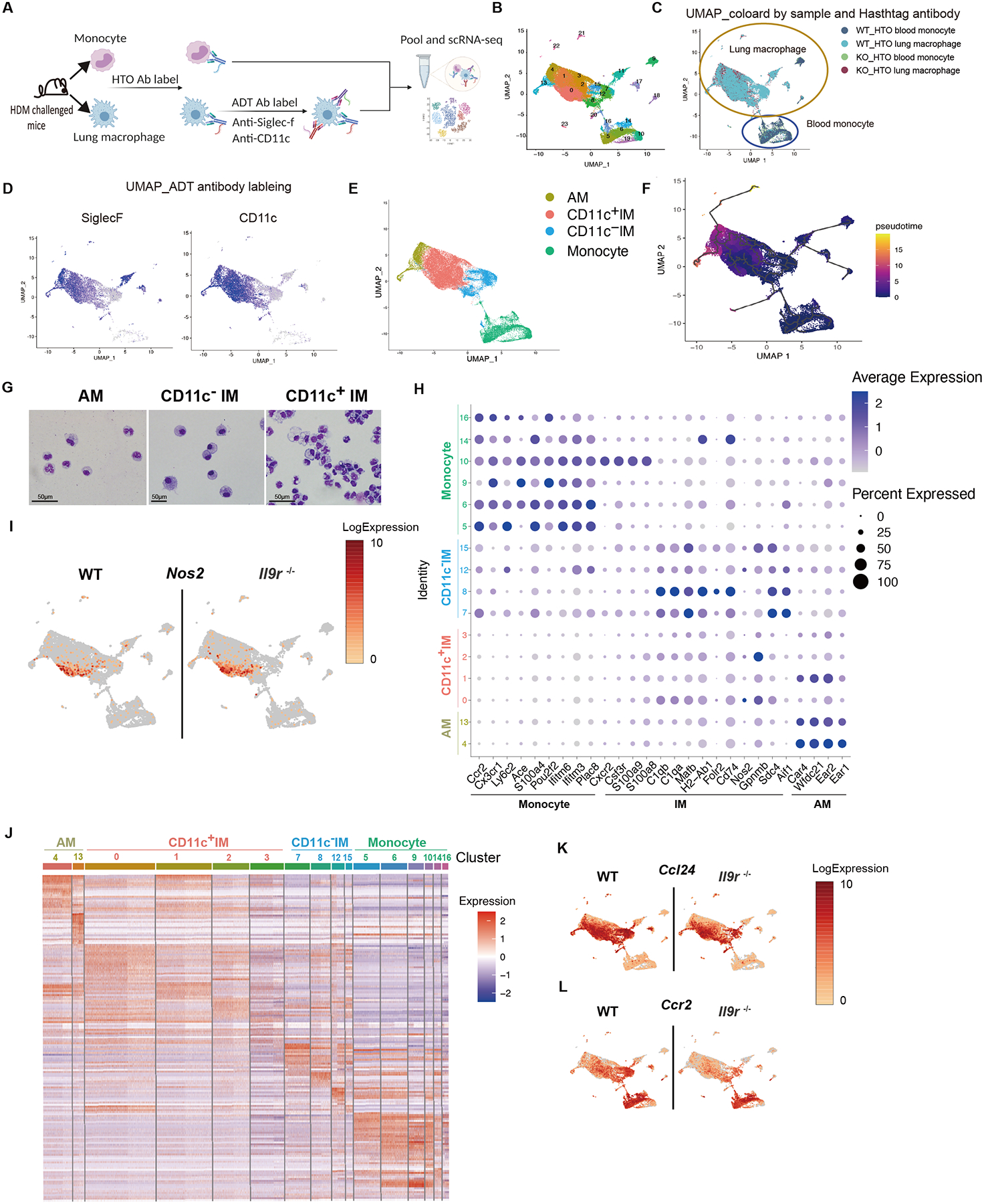
Fig. 3. Heterogeneity of pulmonary macrophages and blood monocytes in allergic inflammation.

(A-G), CITE-seq combined with cell hashing analysis for blood monocytes and lung macrophages from HDM-challenged mice, 15 mice were pooled per group before FACS sorting (A). (B), UMAP showing clusters based on gene expression. (C), UMAP based on cell hashing antibodies. (D), UMAP based on ADT antibodies. (E), Annotation of the lung macrophages within the UMAP based on both transcriptome and surface proteome. (F), Pseudotime analysis using CCR2+ blood monocytes as the root cell type. (G), Lung macrophages were sorted from HDM-treated mice and morphology were analyzed by cytospin.
(H), Dot plot showing the gene expression in different clusters. The number on the y-axis are the cluster numbers shown in (B).
(I), UMAP showing Nos2 expression.
(J), Heatmap showing differential expressed genes in different clusters. The number on the top are the cluster numbers shown in (B).
(K-L), UMAP showing gene expression of the indicated genes.
See also Fig. S3.
