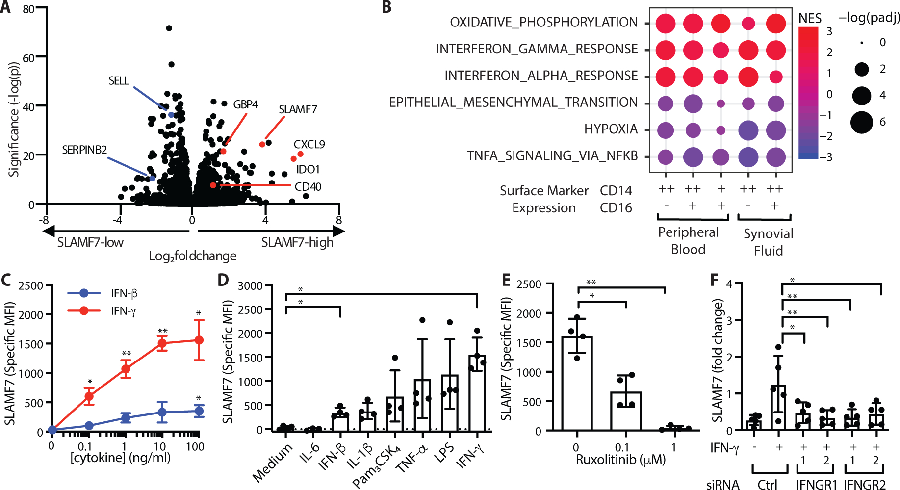
Figure 2. SLAMF7 is a key feature of IFN-γ potentiated macrophages.

A) Differential gene expression in SLAMF7-high versus SLAMF7-low CD14+CD16- cells from peripheral blood (n=12). B) Gene set enrichment analysis for Hallmark Pathways in SLAMF7-high compared to SLAMF7-low myeloid populations from synovial fluid and peripheral blood. C) Specific MFI for SLAMF7 on macrophages incubated with different doses of IFN-γ or IFN-β. D) Specific MFI for SLAMF7 on macrophages incubated with 100 ng/ml of cytokines or TLR agonists. IFN-β and IFN-γ results are the same as the 100 ng/ml dose in panel C. E) Macrophages were incubated with ruxolitinib or DMSO prior to IFN-γ treatment (10 ng/ml), and the specific MFI for SLAMF7 was measured after 16h. Data in C-E represent mean ± SD of 4 donors. F) Macrophages were treated with a siRNA control or siRNA targeting IFNGR1 or IFNGR2, then potentiated with IFN-γ (5–10 ng/ml) for 24h. SLAMF7 was quantified relative to macrophages treated with control siRNA only. Data represent mean ± SD of 5 donors. Statistics were calculated using the one-way ANOVA with Dunnett’s multiple comparisons test. NES, normalized expression score; Ctrl, control; *, p ≤ 0.05; ** p ≤ 0.01.
