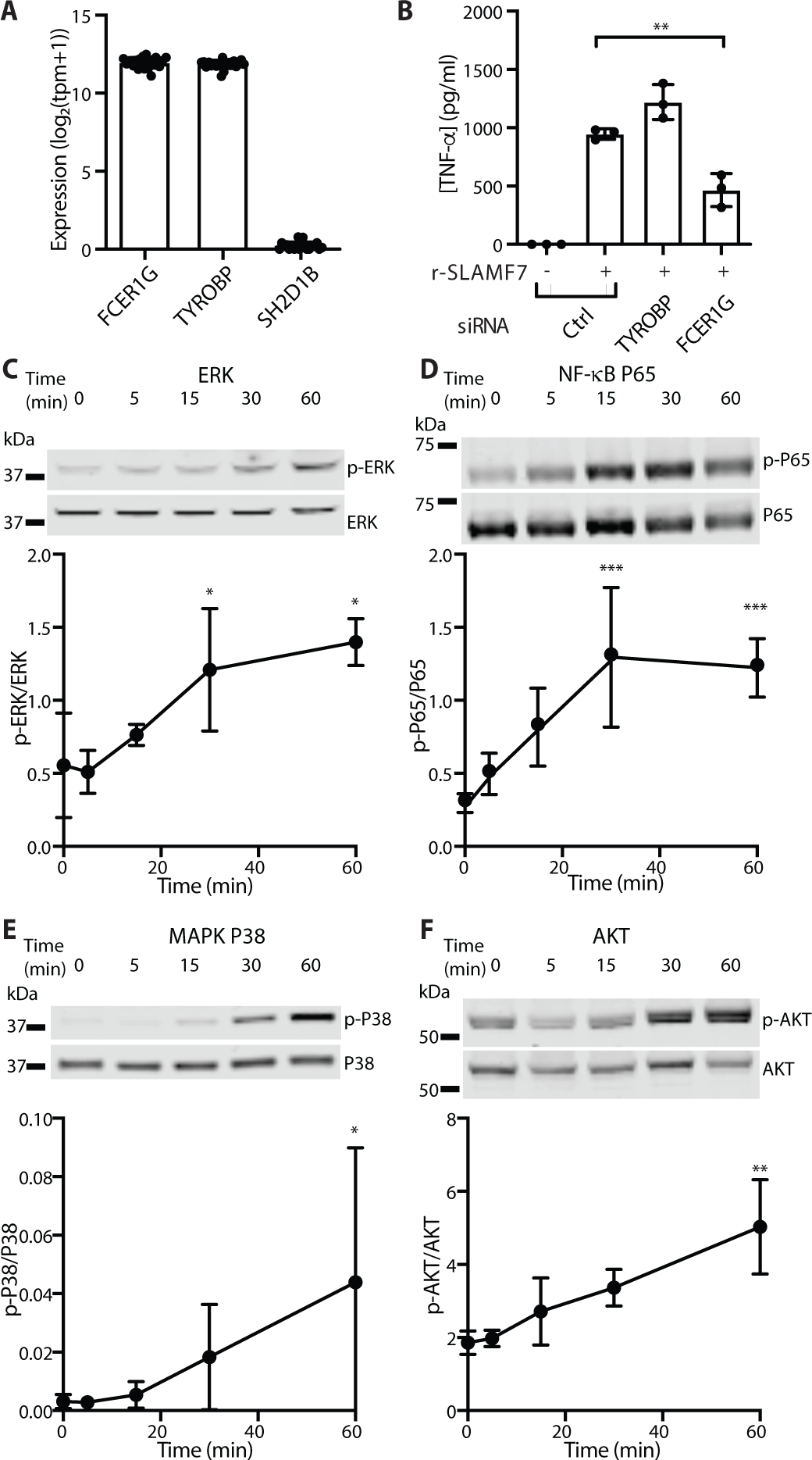
Figure 4. SLAMF7 engagement drives an inflammatory signaling cascade.

A) Gene expression in bulk RNA-seq of synovial tissue macrophages from patients with arthritis from AMP (n=21 donors) (15). B) Macrophages were treated with siRNA control, or siRNA targeting TYROBP or FCER1G. Cells were potentiated with IFN-γ (5 ng/ml) for 24 hours, then stimulated with r-SLAMF7 (100 ng/ml) for 4 hours. Secreted TNF-α was measured by ELISA. Data represent mean ± SD of triplicate wells from an experiment representative of at least 2 independent experiments. C-F) Macrophages were potentiated with IFN-γ (10 ng/ml) for 24h, then stimulated with r-SLAMF7 (100 ng/ml) for the times indicated. Representative Western blots and densitometry quantification for C) ERK and phospho-ERK, D) P65 and phospho-P65, E) MAPK P38 and phospho-MAPK P38, and F) AKT and phospho-AKT. Data represent mean ± SD of at least 3 donors. Statistics were calculated using the one-way ANOVA with Dunnett’s multiple comparisons test. *, p ≤ 0.05; **, p ≤ 0.01; r-SLAMF7, recombinant SLAMF7 protein; tpm, transcripts per million.
