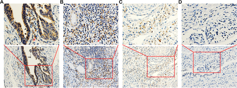
Fig. 1.
Immunohistochemical staining of TIM-1 expression in cervical tissues. A Strong expression of the TIM-1 protein in cervical adenocarcinoma tissue. B Moderate expression of the TIM-1 protein in cervical squamous cell carcinoma tissue. C Weak expression of the TIM-1 protein in cervical intraepithelial neoplasia tissue. D The absence of TIM-1 expression in normal cervical tissue. Lower panels, × 200 magnification; upper panels, × 400 magnification. The red arrowheads indicate positive staining of tumor cells (shown in brown). TIM-1, T-cell immunoglobulin mucin-1

