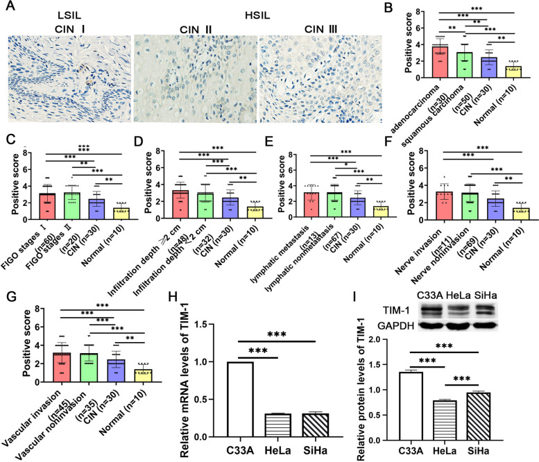
Fig. 2.
The expression of TIM-1 in cervical tissues and CC cell lines. A The expression of the TIM-1 protein in CIN I, II, and III, respectively. B The expression level of TIM-1 in normal cervical, CIN, adenocarcinoma and squamous carinoma. C The expression level of TIM-1 in normal cervical, CIN, FIGO stages I and FIGO stages II. D The expression level of TIM-1 in normal cervical, CIN, Infiltration depth ≥ 1/2 and Infiltration depth < 1/2. E The expression level of TIM-1 in normal cervical, CIN, lymph metastasis and lymph nonmetastasis. F The expression level of TIM-1 in normal cervical, CIN, nerve invasion and nerve noninvasion. G The expression of TIM-1 in normal cervical, CIN, vascular invasion and vascular noninvasion.RT-qPCR (H) and western blotting analysis (I) of the expression of TIM-1 in three CC cell lines (C-33A, Hela, and SiHa). Data are shown as means ± SD. **P < 0.01 and ***P < 0.001. TIM-1, T-cell immunoglobulin mucin-1; CIN, cervical intraepithelial neoplasia; CC, cervical cancer; HSIL, high grade squamous intraepithelial lesion; LSIL, low grade squamous intraepithelial lesion

