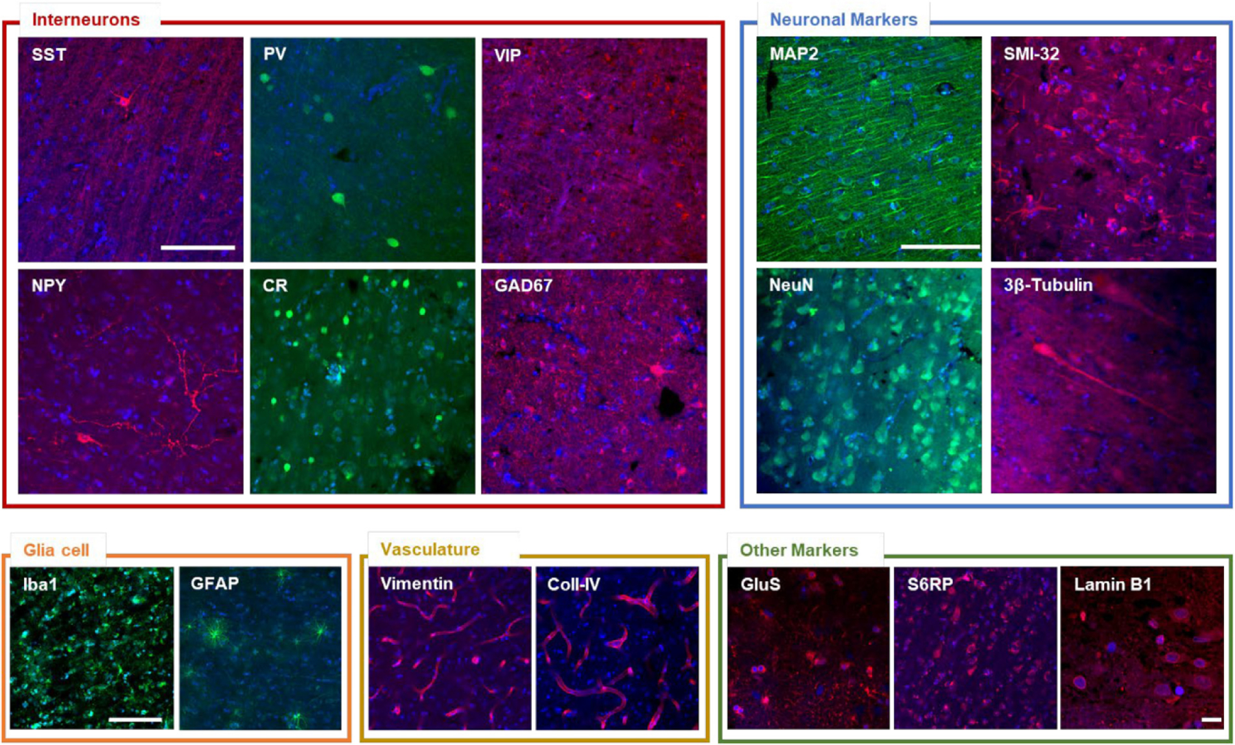
Fig. 3. Antibody validation using confocal microscopy.

Representative confocal images of the different validated staining. Interneurons: somatostatin (SST), parvalbumin (PV), vasointestinal peptide (VIP), neuropeptide Y (NPY), calretinin (CR), glutamic acid decarboxylase (GAD67). Neuronal markers: microtubule-associated protein 2 (MAP2), Non-phosphorylated neurofilament protein (SMI-32 antibody), neuron-specific nuclear protein (NeuN), 3β-Tub (3β-tubulin). Glial cell: ionized calcium binding adaptor molecule 1 (Iba1), glial fibrillary acidic protein (GFAP). Vasculature: vimentin, collagen IV (Coll-IV). Other markers: glutamine synthetase (GluS), phospho-S6 ribosomal protein (S6RP), lamin B1. Pixel size: 0.21 × 0.21 μm. Scale bar = 100 μm. Scale bar for lamin B1 image: 10 μm.
