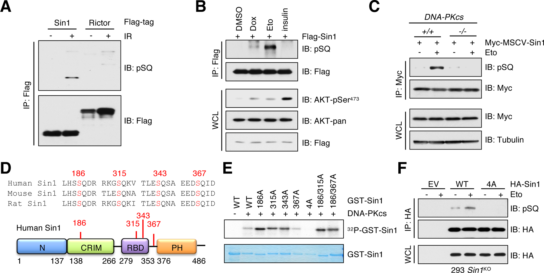
Fig. 2. Sin1 is phosphorylated at SQ motifs by DNA-PK.

(A) IB analysis of Flag immunoprecipitates (IPs) derived from HEK293 cells transfected with Flag-Sin1 or Flag-Rictor. Cells were irradiated with 10 Gy and harvested at 60 min post irradiation (IR). n = 2 independent experiments. (B) IB analysis of whole cell lysates (WCL) and Flag IPs derived from HEK293 cells transfected with Flag-Sin1. Cells were treated with 1 μM doxorubicin (Dox) or 10 μM etoposide (Eto) for 60 min or serum-starved for 12 hours before 100 nM insulin treatment for 30 min. DMSO was used as a control. n = 2 independent experiments. (C) IB analysis of WCL and Myc IPs derived from DNA-PKcs+/+ and DNA-PKcs−/− MEFs stably expressing Myc-Sin1. Cells were treated with 10 μM etoposide for 60 min before harvesting. n = 2 independent experiments. (D) A schematic presentation of the SQ motifs in Sin1. (E) Recombinant GST-Sin1 proteins were purified from E. coli and the active DNA-PKcs was used as the source of kinase in the in vitro kinase assay. Phosphorus-32 (32P) isotope was used to detect phosphorylated Sin1 species. n = 2 independent experiments. (F) IB analysis of WCL and HA IPs derived from HEK293-Sin1KO cells stably expressing HA-Sin1-WT or Sin1–4A. EV (empty vector) was a negative control. Cells were treated with 10 μM etoposide for 60 min before harvesting. n = 3 independent experiments.
