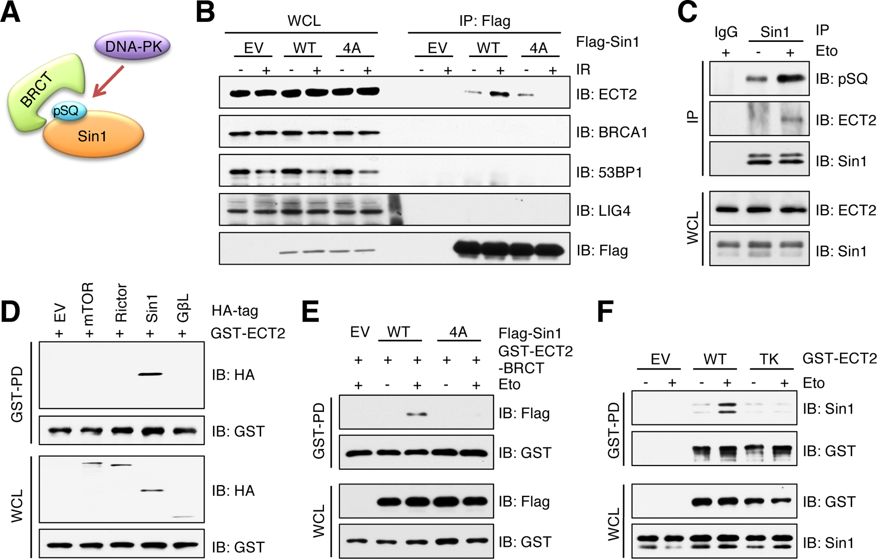
Fig. 4. ECT2 binds Sin1 through BRCT domain-mediated recognition of Sin1-pSQ.

(A) A schematic presentation of hypothesis that BRCT domain interacts with DNA-PK mediated phosphorylation of Sin1-SQ. (B) IB analysis of WCL and Flag IPs derived from HEK293 cells transfected with Flag-Sin1-WT or Sin1–4A. EV was a negative control. Cells were irradiated (IR) with 10 Gy followed by 60 min incubation before harvesting. n = 2 independent experiments. (C) IB analysis of WCL and Sin1 IPs derived from HEK293 cells. IgG was a negative control. Cells were treated with 10 μM etoposide (Eto) for 60 min before harvesting. n = 3 independent experiments. (D) IB analysis of WCL and GST pulldown products (GST-PD) derived from HEK293 cells transfected with indicated HA-tagged constructs and CMV-GST-ECT2. EV was a negative control. n = 2 independent experiments. (E) IB analysis of WCL and GST-PD products derived from MCF7 cells transfected with ECT2-BRCT domains and Sin1-WT or Sin1–4A. EV was a negative control. Cells were treated with 10 μM etoposide for 60 min before harvesting. n = 2 independent experiments. (F) IB analysis of WCL and GST-PD products derived from HEK293 cells transfected with ECT2-WT or ECT2-TK. EV was a negative control. Cells were treated with 10 μM etoposide for 60 min before harvesting. n = 3 independent experiments.
