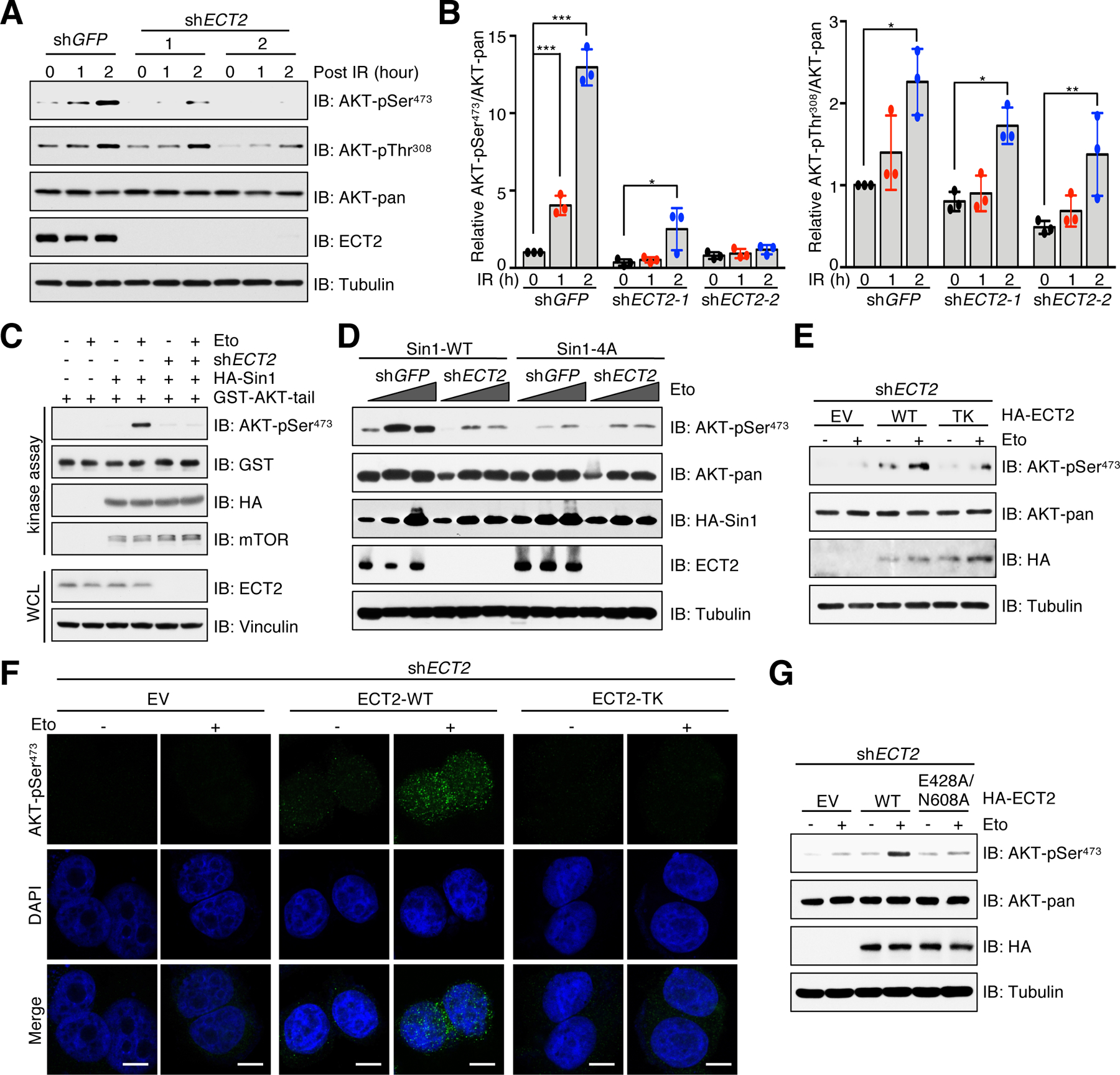
Fig. 5. Functional ECT2 promotes AKT-pSer473 in response to DNA damage.

(A) IB analysis of WCL derived from ECT2-depleted MCF7 cells. shGFP was a negative control. Cells were irradiated (IR) with 10 Gy and incubated for indicated time periods before harvesting. n = 3 independent experiments. (B) Quantification of relative AKT phosphorylation by normalizing phospho-signal to total AKT. Data are means ± SD from three independent experiments. *P < 0.05, **P < 0.01, ***P < 0.001 by one-way ANOVA and Tukey post hoc test. (C) In vitro kinase assays analysis of mTORC2 kinase activity. HA-Sin1 was purified from MCF7-Sin1KO cells that were reconstituted with HA-Sin1 and depleted of ECT2. Cells were treated with 10 μM etoposide (Eto) for 60 min before harvesting. n = 3 independent experiments. (D) MCF7-Sin1KO cells were reconstituted Sin1-WT or Sin1–4A and then infected with ECT2 shRNA. shGFP was a negative control. Cells were treated with 5 or 10 μM etoposide for 60 min before harvesting for IB analysis. n = 3 independent experiments. (E) ECT2-depleted MCF7 cells were reconstituted with EV, ECT-WT or ECT2-TK. Cells were treated with 10 μM etoposide for 60 min before harvesting for IB analysis. n = 3 independent experiments. (F) Cells generated in (E) were treated with 10 μM etoposide for 30 min before performing immunofluorescent staining. Representative images of AKT-pSer473 and DAPI are shown. n = 3 independent experiments. Scale bar, 10 μm. (G) ECT2-depleted MCF7 cells were reconstituted with EV, ECT-WT or ECT2-E428A/N608A. Cells were treated with 10 μM etoposide for 60 min before harvesting for IB analysis. n = 3 independent experiments.
