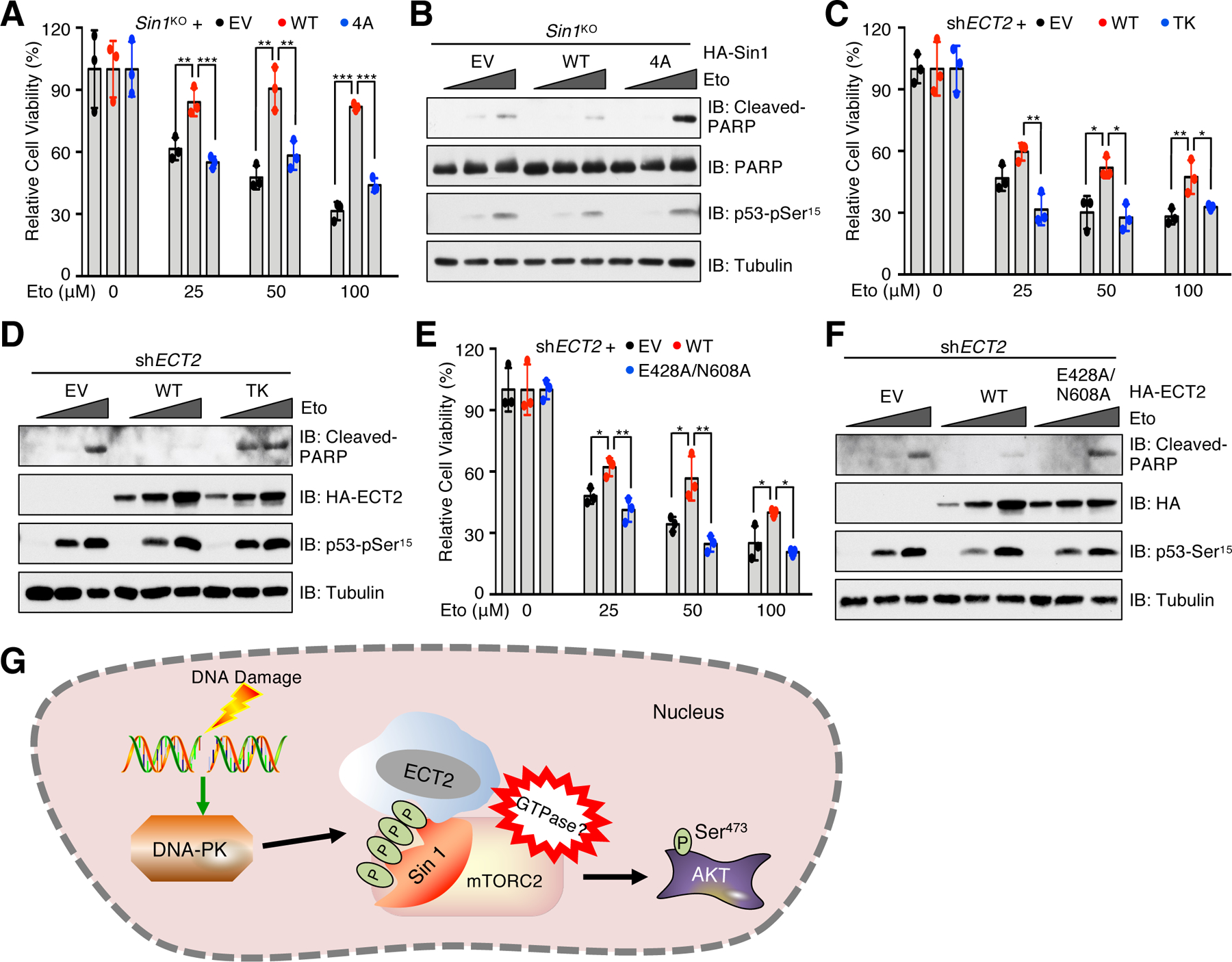
Fig. 6. The Sin1/ECT2 complex promotes cell viability in response to DNA damaging agents.

(A) MCF7-Sin1KO cells reconstituted with EV, Sin1-WT or Sin1–4A were treated with etoposide (Eto) at the indicated doses for 48 hours before examining cell viability. Data are means ± SD from three independent experiments. (B) IB analysis of WCL derived from cells generated in (A). The cells were treated with 0, 50 or 100 μM etoposide for 48 hours before harvesting. n = 3 independent experiments. (C) ECT2-depleted MCF7 cells reconstituted with EV, ECT2-WT or ECT2-TK were treated with etoposide at indicated doses for 48 hours before examining cell viability. Data are means ± SD from three independent experiments. (D) IB analysis of WCL derived from cells generated in (C). The cells were treated with 0, 100 or 300 μM etoposide for 48 hours before harvesting. n = 3 independent experiments. (E) ECT2-depleted MCF7 cells reconstituted with EV, ECT2-WT or ECT2-E428A/N608A were treated with etoposide at indicated doses for 48 hours before examining cell viability. Data are means ± SD from three independent experiments. In (A, C and E), *P < 0.05, **P < 0.01, ***P < 0.001 by one-way ANOVA and Tukey post hoc test. (F) IB analysis of WCL derived from cells generated in (E). The cells were treated with 0, 100 or 300 μM etoposide for 48 hours before harvesting. Blots represent 3 independent experiments. (G) A proposed model illustrating how the DNA-PK/Sin1/ECT2 signaling axis promotes mTORC2 activation to phosphorylate AKT-Ser473 in response to DNA damage.
