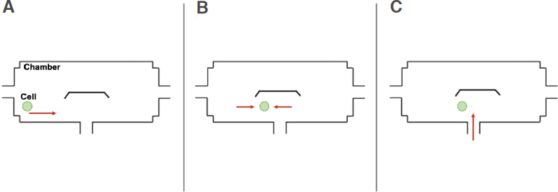
Figure 3.

Schematic diagrams displaying how a single breast cancer cell was sorted and retained near the cell retention structure. A: Cell suspension was inserted through Reservoir 1; B: The liquid level of Reservoirs 1 and 3 was adjusted to make the cell stop near the cell retention structure; C: Upon cell retention, chemical reagents were administrated through Reservoir 2
