
Figure 1. Representative Photomicrographs of Human Hippocampi Depicting the Main Neuropathologic Features Analyzed in the Current Study: LATE-NC + Pathology Without (A and B) or With (C and D) Comorbid HS Pathology.
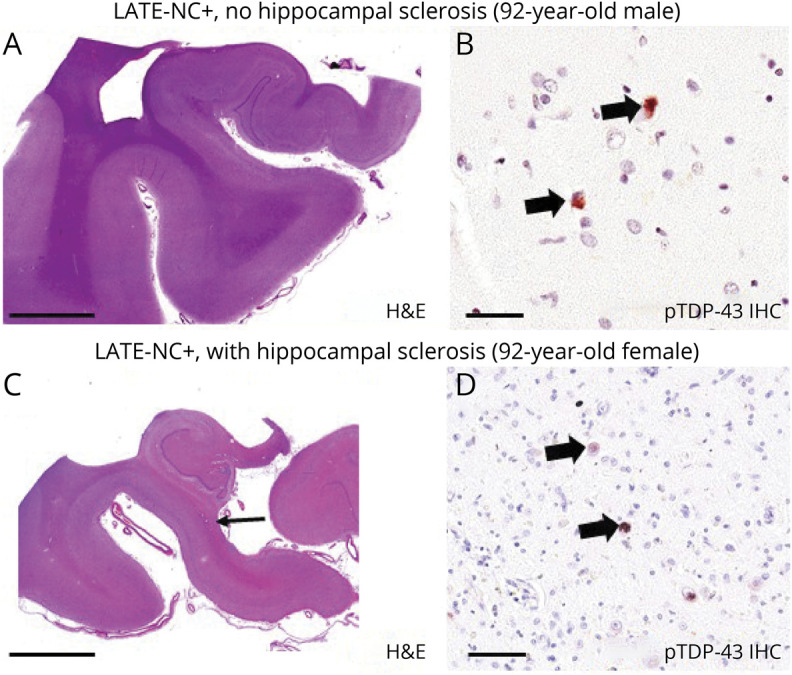
Each photomicrograph depicts anterior hippocampi dissected in the coronal plane. (A and B) Hematoxylin & eosin (H&E) and phospho (p)–TAR DNA binding protein 43 (TDP-43) immunohistochemistry (IHC), respectively, from a man who died at 92 years of age. Autopsy revealed limbic-predominant age-related TDP-43 encephalopathy neuropathologic changes (LATE-NC) stage 2 but no hippocampal sclerosis (HS) pathology. (C and D) Results from a woman who also died at 92 years of age. In her case, the autopsy revealed comorbid LATE-NC stage 2 and HS pathology. Note the relatively fulsome hippocampal profile in panel A compared to panel C (same scale bar); the HS+ profile in panel C shows thinning in CA1 and subiculum (arrow). Higher-magnification assessment confirmed that there was substantial neuronal cell dropout and robust astrocytosis (not shown). pTDP-43–positive intraneuronal inclusions are highlighted with arrows in panels B and D. These representative photomicrographs were from research participants of the University of Kentucky Alzheimer’s Disease Research Center. Scale bar = 2 mm in panels A and C, 70 μm in panel B, and 100 μm in panel D.