
Fig 4. Comparison of HTT MSD assays employing only monoclonal antibodies (right-hand panels) with existing assays using polyclonal antibodies (left-had panels).
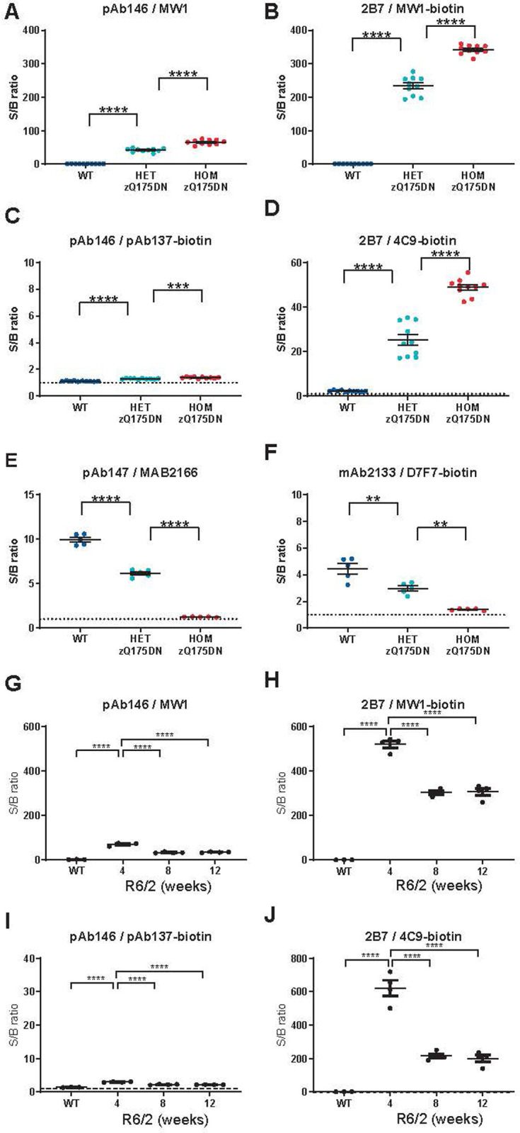
Panels show gene dosage effect in wild type, heterozygous and homozygous zQ175DN knock-in mouse striatal lysates (A-D) and zQ175DN mouse brain hemisphere lysates (E, F), and age-related HTT levels in wild type and R6/2 mouse brain hemisphere lysates (G-J). Dissected snap-frozen tissues were lysed and analyzed with HTT MSD assays employing the indicated antibody combinations for capture/detection of polyglutamine-expanded HTT (A,B,G,H), total human HTT (C,D,I,J) or mouse HTT (E,F). Each data point shows the average of n = 3 technical replicates; overlays show mean ± SEM by genotype (Q175 n = 10 per group; Q175DN n = 5 per group; R6/2 n = 4 per group). Data are shown as MSD signal over background ratio. Data in A–F were analyzed with unpaired t-tests with Bonferroni correction, data in G-J with Dunnet’s post hoc test following ANOVA. ****p < 0.0001; ***p < 0.001, **p < 0.01.