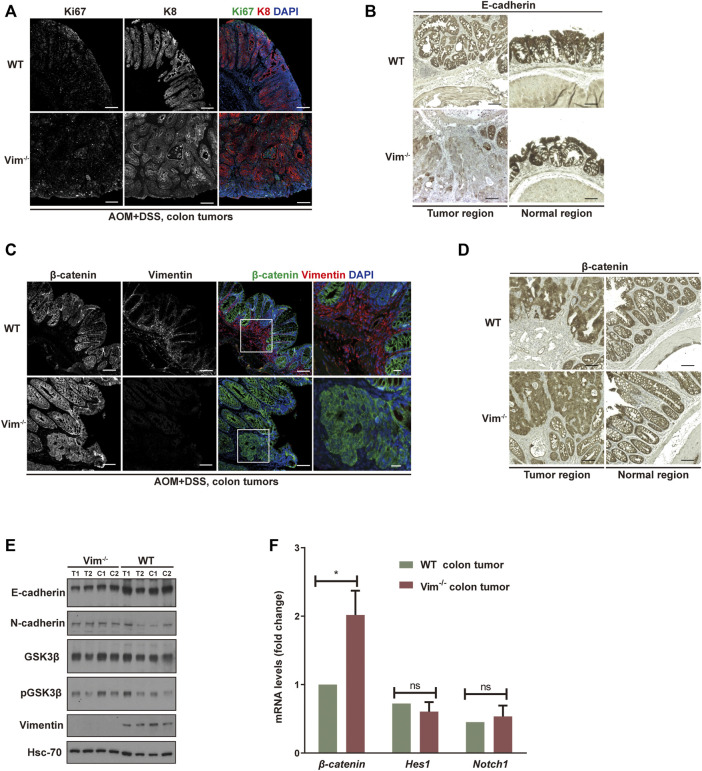
FIGURE 4.
Increased proliferation and tumor grade in Vim−/− cancer. (A) Representative confocal images indicated the expression of ki67 (in green), K8 (in red), and DAPI (in blue) in WT and Vim−/− colon tumors upon AOM and DSS treatment. (B) Representative pictures of tumors and their neighboring normal regions by immunohistochemical labeling of E-cadherin of WT and Vim−/− mouse colon upon AOM + DSS induction. (C) Representative confocal images of the expression of β-catenin (in green), vimentin (in red), and DAPI (in blue) in WT and Vim−/− colon tumors upon AOM and DSS treatment. (D) Representative pictures of tumors and their neighboring normal regions by immunohistochemical labeling of β-catenin of WT and Vim−/− mouse colon upon AOM + DSS induction. Scale bar (A–D), 200 μm. (E) Extracts (30 μg) from colon tumors of WT and Vim−/− mice were immunoblotted with anti–E-cadherin, anti–N-cadherin, anti–GSK-3β, anti–P-GSK-3β, or anti-vimentin. Hsc-70 blotted from the lysates to control for equal loading. (F) Quantitative real-time PCR (qRT-PCR) analysis of transcripts for β-catenin, Hes-1, and Notch-1 in WT and Vim−/− mouse colon tumors. Error bars = ± SEM; n =6; *, p < 0.05; ns, not significant.

