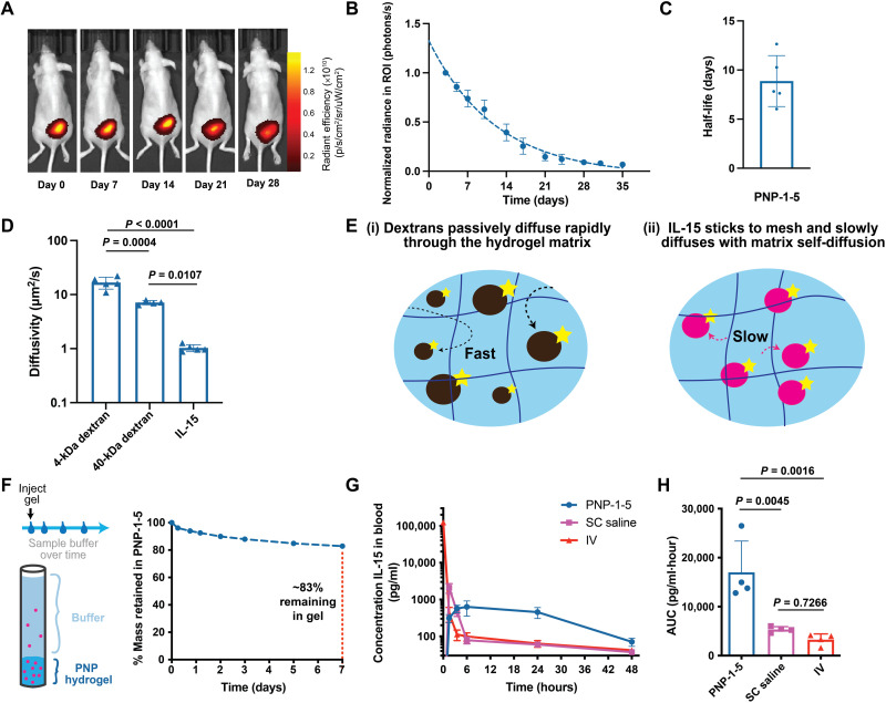
Fig. 3. PNP hydrogels enable prolonged retention of stimulatory cytokines and exhibit controlled degradation in vivo.
(A) In vivo imaging of fluorescently tagged PNP-1-5 hydrogel over 4 weeks after subcutaneous injection in mice. (B) Quantification of fluorescence in the region of interest (ROI) surrounding the hydrogel and accompanying representative images over time. (C) Degradation half-life of PNP-1-5 hydrogels in vivo. (D) Diffusivity of FITC-dextran molecules and FITC-labeled IL-15 in the PNP-1-5 hydrogel formulation as measured by fluorescence recovery after photobleaching. (E) Schematic illustrating the different diffusion mechanisms of the (i) dextran molecules and (ii) IL-15 in the PNP hydrogel mesh. (F) Schematic of in vitro release assay of IL-15 from PNP-1-5 hydrogel immersed in saline over 1 week. %Mass of IL-15 remaining in the PNP-1-5 hydrogel during the release assay. (G) Pharmacokinetic curves of IL-15 in the blood administered intravenously, subcutaneously (SC), and from PNP-1-5 injected subcutaneously in mice. (H) Area under the curve (AUC) of the pharmacokinetic profiles of the various IL-15 administration routes.

