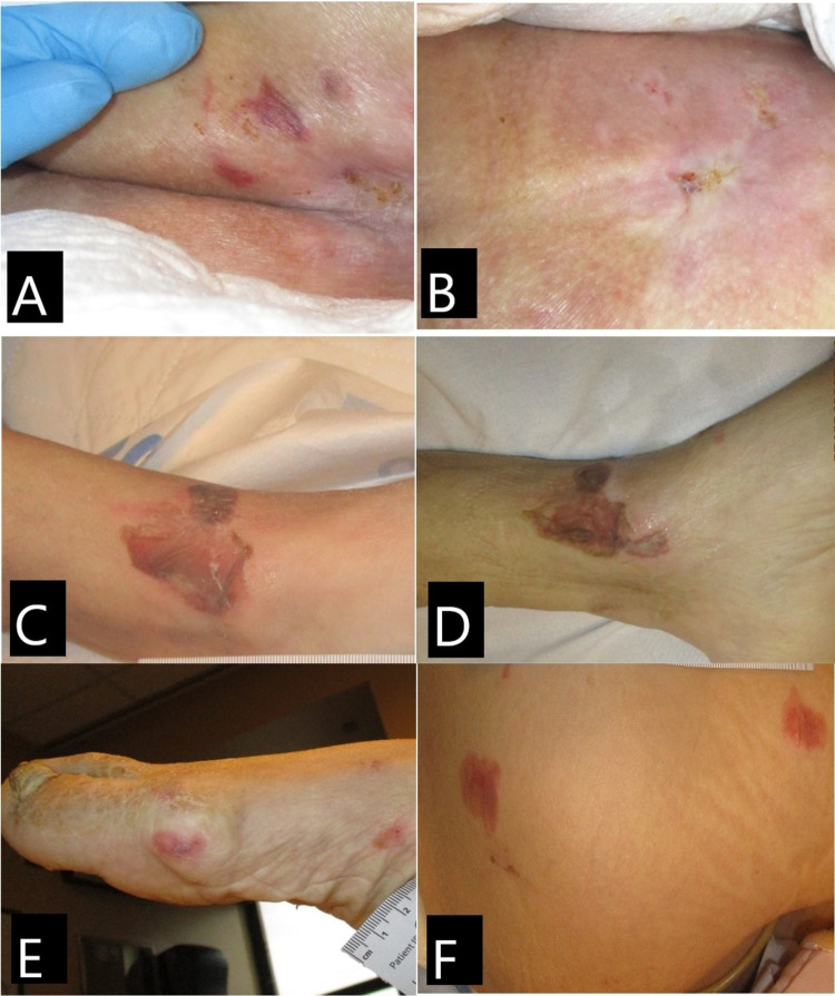
Figure 1. Case 1. Skin lesions.
(A) Buttocks on day 1: purpuric patch, nonblanchable. (B) Buttocks on day 10: lesions resolved; note whitish atrophic appearance of the recent healed skin. (C) Left ankle of day 1: serosanguineous-filled bullae. (D) Left ankle on day 10: bullae with no drainage. (E) Right foot on day 1: purpuric nonblanchable patch. (F) Right lateral thigh: nonblanchable purpuric patches and macules.

